Case Reports
. 2017 Oct;37(10):1820-1831.
doi: 10.1097/IAE.0000000000001448.

PNEUMATIC VITREOLYSIS FOR RELIEF OF VITREOMACULAR TRACTION

Affiliations
Case Reports

PNEUMATIC VITREOLYSIS FOR RELIEF OF VITREOMACULAR TRACTION

Clement K Chan et al. Retina. 2017 Oct.

Abstract

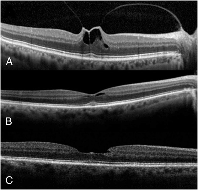
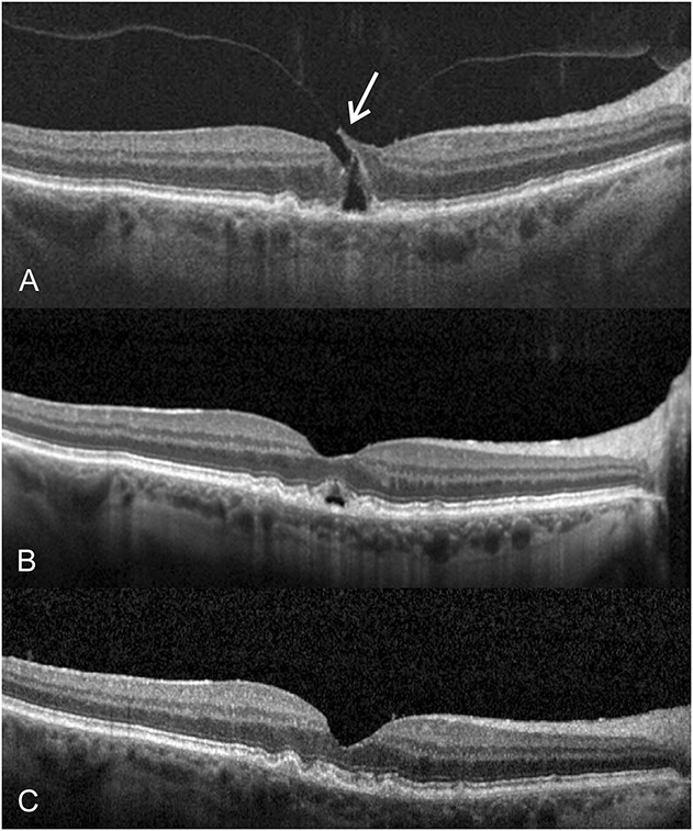
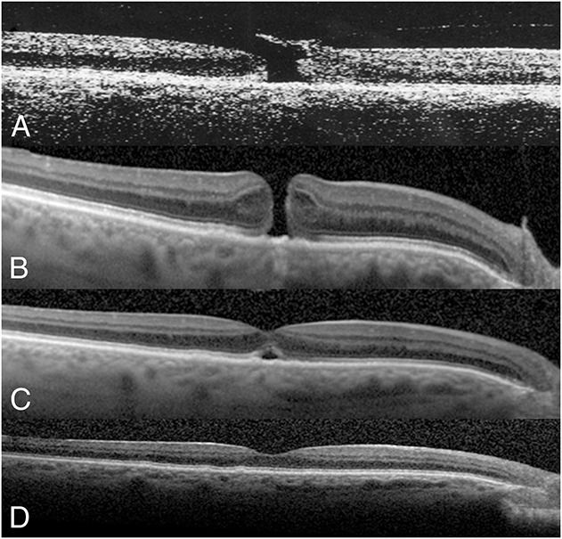
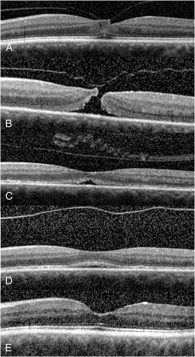
Purpose: To evaluate the outcome of perfluoropropane (C3F8) gas injection for symptomatic vitreomacular traction (VMT) with or without Stage 2 macular hole (MH).

Methods: A retrospective review of eyes with VMT treated with 0.3 mL of C3F8 gas was performed. Patients avoided the supine position until gas resolution. Patients with small MH maintained partial face-down positioning.

Results: Forty-nine consecutive patients (50 eyes) with symptomatic VMT underwent pneumatic vitreolysis between 2010 and 2016. A posterior vitreous detachment developed in 43 eyes (86.0%) after a single gas injection, at a median of 3.0 weeks. Twenty-eight of 35 eyes (80.0%) with VMT only and all 15 eyes (100%) with a small Stage 2 MH developed a posterior vitreous detachment, with MH closure in 10 of 15 eyes (66.7%). Median baseline and last best spectacle-corrected visual acuities were 20/50 and 20/40, respectively (P < 0.001). Mean follow-up time was 11.1 ± 9.9 months. Rate of posterior vitreous detachment was reduced with presence of diabetes mellitus (25%) and with thick cellophane membrane (50%). Univariate analysis showed increased VMT release for eyes with VMT extent within 1 disk area (χ = 13.1, P = 0.002), eyes with absence of diabetes mellitus (χ = 8.8, P = 0.007), and eyes with Stage 2 MH (χ = 5.47, P = 0.019); there was a trend between success and lack of thick cellophane membrane (χ = 3.32, P = 0.068). Results using logistic regression also showed younger age (P = 0.012), followed by better baseline best spectacle-corrected visual acuity (P = 0.044), lack of diabetes mellitus (P = 0.077), and female gender (P = 0.045) to be predictors of increased VMT release. One VMT-only eye formed a MH and another VMT-only eye developed a retinal detachment. Both eyes responded to vitrectomy.

Conclusion: Pneumatic vitreolysis with limited face-down position is a viable option for treating VMT with few adverse events. More studies are needed to elucidate its indications, benefits, and risks.


Conflict of interest statement

None of the authors have any financial/conflicting interests to disclose.

Figures

Fig. 1.
Fig. 1.
A 72-year-old woman presented with a complaint of blurred vision and metamorphopsia involving her right eye. The BSCVA was 20/40 in the right eye, and there was central VMT of slightly more than 1 DA in size on SD-OCT (A). At 5 weeks after injection of 0.3 mL of C3F8 gas, a PVD developed and the VMT was relieved, but there was mild residual perifoveal cystic lesion in the right eye (B). At 18 months after PVL, there was a normal macular appearance and the BSCVA was 20/30 in the right eye (C).
Fig. 2.
Fig. 2.
A 71-year-old man initially presented with asymptomatic vitreomacular adhesion without a macular defect in his right eye in September 2013. The BSCVA was 20/25 in the right eye. Six months later, he returned with a complaint of blurred and distorted vision in the right eye. His BSCVA had deteriorated to 20/70 in the right eye, and SD-OCT showed central VMT associated with a narrow Stage 2 MH with a retinal flap (arrow) in the right eye (A). At 1 week after injection of 0.3 mL of C3F8 gas and partial face-down positioning, a PVD developed with a partial resolution of the MH. However, there was focal outer foveal lucency, corresponding to a residual outer foveal defect noted on SD-OCT (B). Subsequent visits showed progressive resolution of the outer foveal defect. The BSCVA was improved to 20/40, corresponding to a complete closure of the MH, in the right eye at 5 months after gas injection (C).
Fig. 3.
Fig. 3.
A 72-year-old woman presented with VMT associated with a small Stage 2 MH in the right eye (A). The BSCVA was 20/50 in the right eye. After electing PVL for treatment of the VMT, she received 0.3 mL of C3F8 gas in the right eye. Despite relief of VMT and partial narrowing of the foveal defect within 8 days after PVL, the MH did not close (B). She declined a vitrectomy and decided on receiving a second C3F8 gas bubble instead, which was performed for right eye without complications at 10 days later. She maintained face-down positioning and there was closure of inner layers of MH at 4 days after injection of the second gas bubble in the right eye (C). There was further closure of the outer foveal defect in subsequent weeks. At 6 months after surgery, there was complete closure of MH with BSCVA of 20/30 in the right eye (D).
Fig. 4.
Fig. 4.
A 67-year-old man complained of progressive central visual deficit of his left eye in August 2013. His BSCVA was 20/70 in the left eye. The SD-OCT showed central vitreomacular adhesion only without symptoms in his right eye, but symptomatic central VMT associated with a partial split of the foveal layers, consistent with a Stage 1 impending MH in the left eye (A). He elected to undergo ocriplasmin injection. Within 24 hours after ocriplasmin injection, he reported further visual loss and an urgent examination showed residual VMT and the progressing of the Stage 1 impending MH to a Stage 2 full-thickness MH with residual VMT in the right eye (B). The BSCVA was deteriorated to 20/100 in the right eye. He then elected to undergo PVL. At 4 days after receiving 0.3 mL of C3F8 gas injection and face-down positioning, a PVD with closure of the inner layers of the MH developed (C). There was a partial lucency of the outer fovea, corresponding to the residual outer foveal defect, in the right eye. At 4 weeks after PVL, there was a decrease in the outer foveal defect (D). At 6 weeks after PVL, there was a complete closure of the MH with BSCVA recovery to 20/30 in the left eye (E).

References

    1. Sebag J. Is pharmacologic vitreolysis brewing? Retina 2002;22:1–3. - PubMed
    1. Sebag J. Pharmacologic vitreolysis, premise and promise of the first decade. Retina 2009;29:871–874. - PubMed
    1. Duker JS, Kaiser PK, Binder S, et al. The International Vitreomacular Traction Study Group classification of vitreomacular adhesion, traction, and macular hole. Ophthalmology 2013;120:2611–2619. - PubMed
    1. Hikichi T, Yoshida A, Trempe CL. Course of vitreomacular traction syndrome. Am J Ophthalmol 1995;119:55–61. - PubMed
    1. Sulkes DJ, Ip MS, Baumal CR, et al. Spontaneous resolution of vitreomacular traction documented by optical coherence tomography. Arch Ophthalmol 2000;118:286–287. - PubMed

Publication types

MeSH terms