Atorvastatin-induced necrotizing autoimmune myositis: An emerging dominant entity in patients with autoimmune myositis presenting with a pure polymyositis phenotype
- PMID: 28099331
- PMCID: PMC5279076
- DOI: 10.1097/MD.0000000000005694
Atorvastatin-induced necrotizing autoimmune myositis: An emerging dominant entity in patients with autoimmune myositis presenting with a pure polymyositis phenotype
Abstract
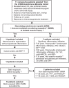
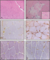
The general aim of this study was to evaluate the disease spectrum in patients presenting with a pure polymyositis (pPM) phenotype. Specific objectives were to characterize clinical features, autoantibodies (aAbs), and membrane attack complex (MAC) in muscle biopsies of patients with treatment-responsive, statin-exposed necrotizing autoimmune myositis (NAM). Patients from the Centre hospitalier de l'Université de Montréal autoimmune myositis (AIM) Cohort with a pPM phenotype, response to immunosuppression, and follow-up ≥3 years were included. Of 17 consecutive patients with pPM, 14 patients had a NAM, of whom 12 were previously exposed to atorvastatin (mean 38.8 months). These 12 patients were therefore suspected of atorvastatin-induced AIM (atorAIM) and selected for study. All had aAbs to 3-hydroxy-3-methylglutaryl coenzyme A reductase, and none had overlap aAbs, aAbs to signal recognition particle, or cancer. Three stages of myopathy were recognized: stage 1 (isolated serum creatine kinase [CK] elevation), stage 2 (CK elevation, normal strength, and abnormal electromyogram [EMG]), and stage 3 (CK elevation, proximal weakness, and abnormal EMG). At diagnosis, 10/12 (83%) patients had stage 3 myopathy (mean CK elevation: 7247 U/L). The presenting mode was stage 1 in 6 patients (50%) (mean CK elevation: 1540 U/L), all of whom progressed to stage 3 (mean delay: 37 months) despite atorvastatin discontinuation. MAC deposition was observed in all muscle biopsies (isolated sarcolemmal deposition on non-necrotic fibers, isolated granular deposition on endomysial capillaries, or mixed pattern). Oral corticosteroids alone failed to normalize CKs and induce remission. Ten patients (83%) received intravenous immune globulin (IVIG) as part of an induction regimen. Of 10 patients with ≥1 year remission on stable maintenance therapy, IVIG was needed in 50%, either with methotrexate (MTX) monotherapy or combination immunosuppression. In the remaining patients, MTX monotherapy or combination therapy maintained remission without IVIG. AtorAIM emerged as the dominant entity in patients with a pPM phenotype and treatment-responsive myopathy. Isolated CK elevation was the mode of presentation of atorAIM. The new onset of isolated CK elevation on atorvastatin and persistent CK elevation on statin discontinuation should raise early suspicion for atorAIM. Statin-induced AIM should be included in the differential diagnosis of asymptomatic hyperCKemia. Three patterns of MAC deposition, while nonpathognomonic, were pathological clues to atorAIM. AtorAIM was uniformly corticosteroid resistant but responsive to IVIG as induction and maintenance therapy.
Conflict of interest statement
Authors YT, OL-C, JF, ER, MG, J-RG, JB-T, YR, JD, AA, and J-LS have no conflicts of interest to disclose. The authors listed below have received financial support (personal or institutional) from the listed institutions, unrelated to the present work. INT: Oklahoma Medical Research Foundation Clinical Immunology Laboratory, UpToDate; MJF: EUROIMMUN, ImmunoConcepts, Inova Diagnostics.
Figures


References
-
- Bohan A, Peter JB. Polymyositis and dermatomyositis (parts 1 and 2). N Engl J Med 1975;292:344–7. 403–407. - PubMed
-
- Troyanov Y, Targoff I, Tremblay JL, et al. Novel classification of idiopathic inflammatory myopathies based on overlap clinical features and autoantibodies: analysis of 100 French Canadian patients with myositis. Medicine (Baltimore) 2005;84:231–49. - PubMed
-
- van de Vlekkert J, Hoogendijk JE, de Visser M. Myositis with endomysial cell invasion indicates inclusion body myositis even if other criteria are not fulfilled. Neuromusc Disord 2015;25:451–6. - PubMed
-
- van der Meulen MF, Bronner IM, Hoogendijk JE, et al. Polymyositis: an overdiagnosed entity. Neurology 2003;61:316–21. - PubMed
-
- Dalakas MC. Inflammatory muscle diseases. N Engl J Med 2015;372:1734–47. - PubMed
Publication types
MeSH terms
Substances
LinkOut - more resources
Full Text Sources
Other Literature Sources
Medical
Research Materials

