. 2017 Jan 26;541(7638):488-493.
doi: 10.1038/nature21049. Epub 2017 Jan 18.

Communication between viruses guides lysis-lysogeny decisions

Affiliations

Communication between viruses guides lysis-lysogeny decisions

Zohar Erez et al. Nature. .

Abstract

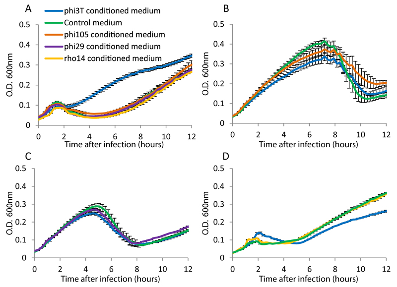
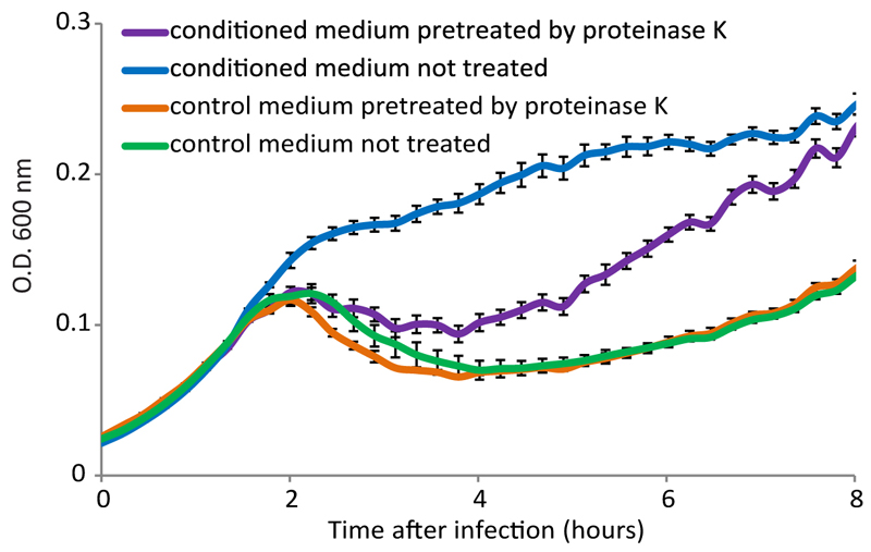
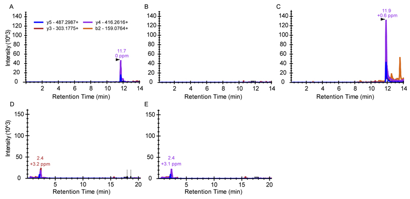
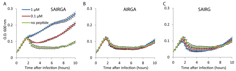
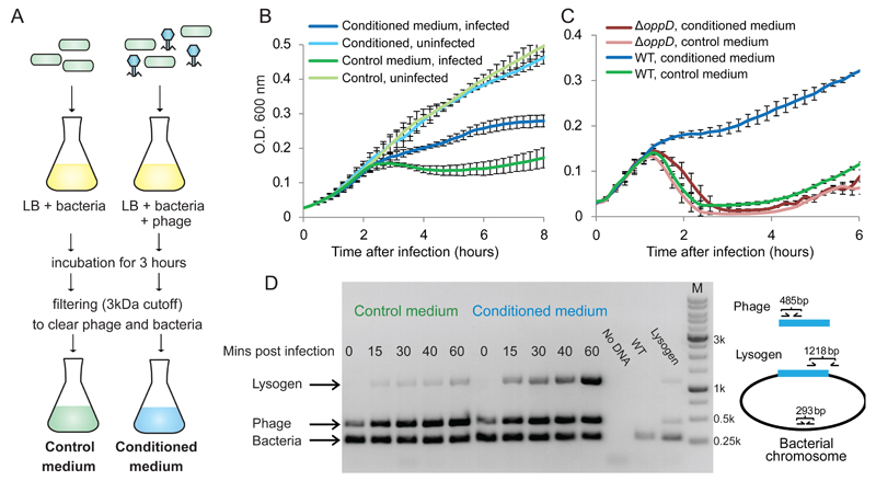
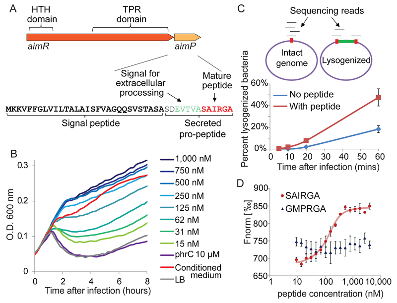
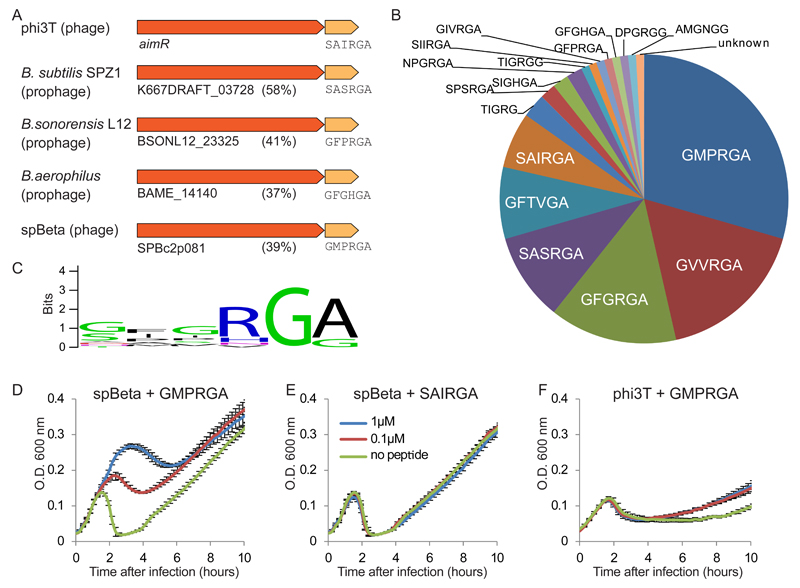
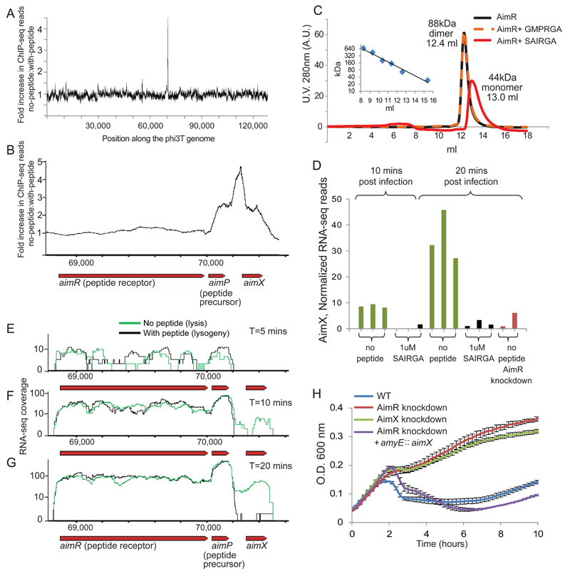
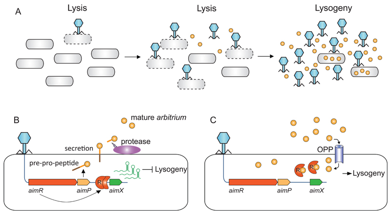
Temperate viruses can become dormant in their host cells, a process called lysogeny. In every infection, such viruses decide between the lytic and the lysogenic cycles, that is, whether to replicate and lyse their host or to lysogenize and keep the host viable. Here we show that viruses (phages) of the SPbeta group use a small-molecule communication system to coordinate lysis-lysogeny decisions. During infection of its Bacillus host cell, the phage produces a six amino-acids-long communication peptide that is released into the medium. In subsequent infections, progeny phages measure the concentration of this peptide and lysogenize if the concentration is sufficiently high. We found that different phages encode different versions of the communication peptide, demonstrating a phage-specific peptide communication code for lysogeny decisions. We term this communication system the 'arbitrium' system, and further show that it is encoded by three phage genes: aimP, which produces the peptide; aimR, the intracellular peptide receptor; and aimX, a negative regulator of lysogeny. The arbitrium system enables a descendant phage to 'communicate' with its predecessors, that is, to estimate the amount of recent previous infections and hence decide whether to employ the lytic or lysogenic cycle.


Figures

Extended data figure 1
Extended data figure 1. Specificity of conditioned media.
Conditioned media were prepared using initial infection of B. subtilis 168 by four phages: phi3T, phi105, phi29 and rho14. Presented are growth curves of B. subtilis 168 in the different media, infected with each phage at MOI=0.1. (A) Infection with phi3T. (B) Infection with phi105. (C) Infection with phi29. (D) Infection with rho14. Data represent average of 3 replicates, and error bars represent standard error.
Extended data figure 2
Extended data figure 2. Proteinase K treatment reduces the effect of conditioned medium.
Growth curves of B. subtilis 168 infected by phi3T at MOI=0.1, in control and conditioned media, with and without pre-treatment with proteinase K. Data represent average of 3 technical replicates, and error bars represent standard error.
Extended data figure 3
Extended data figure 3. Mass spectrometry verifies the presence of the SAIRGA peptide in conditioned medium.
Presented are extracted ion chromatograms of targeted MS analysis experiment. B2, Y3, Y4 and Y5 refer to fragmentation products of the peptides made by the instrument in the MS/MS process, with the expected m/z ion masses indicated. Y-axis represents the ion intensity of each fragment ion (arbitrary units). (A) Reference synthesized SAIRGA peptide at 100 nM concentration in LB. (B) Control medium from B. subtilis 168. (C) Conditioned medium from phi3T-infected B. subtilis 168. Arrowhead depicts the expected retention time of the SAIRGA peptide. (D) Control medium from B. subtilis 168 expressing dCas9 with a spacer targeting aimP. (F) Conditioned medium derived from phi3T-infected B. subtilis 168 expressing dCas9 with a spacer targeting aimP.
Extended data figure 4
Extended data figure 4. 5aa versions of the 6aa arbitrium peptide do not guide lysogeny.
Growth curves of B. subtilis 168 infected by phi3T at MOI=0.1, in LB media supplemented with synthesized SAIRGA, AIRGA or SAIRG peptide. Shown is average of 3 biological replicates, each with 3 technical replicates. Error bars represent standard error.
Extended data figure 5
Extended data figure 5. Exposure of AimR to SAIRGA peptide reduces propensity for dimerization.
Purified AimR was eluted from a gel-filtration column, dialyzed, then mixed with SAIRGA peptide (f.c. 100 µM) dissolved in water or with equal amount of volume without peptide and incubated at room temperature for 5 minutes. The protein samples were then mixed for 30 minutes with different concentrations of the crosslinker BS3 bis(sulfosuccinimidyl)suberate. Presented are electrophoresis results analyzed using the TapeStation instrument (Agilent Technologies), showing that in the absence of peptide, purified AimR tends to preferentially be cross linked into dimers, whereas in the presence of the SAIRGA peptide, dimerization is significantly reduced.
Extended data figure 6
Extended data figure 6. Phage gene expression 20 minutes post infection.
Each dot represents a single phage gene. Axes represent average RNA-seq read count per gene from 3 replicates, after normalization to control for RNA-seq library size. X axis, expression when phage infection was in the presence of 1 µM of SAIRGA peptide; Y axis, no peptide.
Extended data figure 7
Extended data figure 7. Expression of the arbitrium locus in phage spBeta during infection.
RNA-seq coverage of the arbitrium locus at 20 minutes post infection, with (black) or without (green) 1 µM of synthesized GMPRGA peptide in the medium. RNA-seq coverage was normalized to the number of reads mapped to the phage genome in each RNA-seq library. Shown are two replicates of the experiment.
Extended data figure 8
Extended data figure 8. Infection experiments with phi3T delta-aimP and derived conditioned media.
Growth curves of B. subtilis 168 infected with phi3T or phi3T delta-aimP in (A) conditioned medium derived from phi3T; (B) conditioned medium derived from phi3T delta-aimP; and (C) control medium. Each medium was generated in biological replicate, and the two replicates are presented separately. Each curve represents the average of three technical replicates, and error bars represent standard error.
Figure 1
Figure 1. Effect of conditioned media on the infection dynamics of phage phi3T.
(A) Preparation protocol of control and conditioned media. (B) Growth curves of B. subtilis 168 infected by phi3T at MOI=0.1, in control and conditioned media. (C) Growth curves of B. subtilis strain 3610 (WT) and its derivative DS4979 (oppD::kan) infected by phi3T at MOI=0.1. For panels B-C, data represents average of 3 biological replicates, each with 3 technical replicates; error bars represent SE. (D) Semi-quantitative PCR assay for phage lysogeny during an infection time course of B. subtilis 168 with phi3T. “No DNA”, control without DNA; “WT”, DNA from uninfected culture; “Lysogen”, genomic DNA of a phi3T lysogen .
Figure 2
Figure 2. The arbitrium peptide and its receptor.
(A) The arbitrium locus in the phage genome. (B) Growth curves of B. subtilis 168 infected by phi3T at MOI=0.1, in LB media supplemented with synthesized SAIRGA peptide. Numbers represent peptide concentrations. Average of 3 biological replicates, each with 3 technical replicates. (C) Sequencing-based quantitative determination of the fraction of lysogenized bacteria. Top – schematics of the analysis. Percent lysogeny was calculated as the fraction of reads spanning the phage/bacteria integration junction out of total reads covering this junction. Integration junction is red; integrated phage is green. Bottom: percent lysogenized bacteria during infection of B. subtilis 168 with phi3T at MOI=2. Average of three biological replicates, error bars denote SE. Synthesized SAIRGA peptide was added at 1 µM. (D) Microscale thermophoresis (MST) analysis of the binding between purified AimR and synthesized SAIRGA or GMPRGA peptides. Average and SE of three replicates.
Figure 3
Figure 3. Accumulation of arbitrium peptide during an infection time course.
B. subtilis 168 culture was grown to OD=0.5 and then infected at t=0 by phi3T at MOI=0.001. PFU, plaque forming units; CFU, colony forming units, sampled from the uninfected control. For peptide, PFU and OD shown is average of triplicate (except for t=30 where it is duplicate) with error bars representing SE. CFU was measured in duplicates, and error bars represent the two measured points around the average. (A) Growth of infected and uninfected bacteria, and phage, during the infection time course. (B) Accumulation of peptide during the infection course as compared to bacteria and phage.
Figure 4
Figure 4. A peptide communication code guiding lysogeny in Bacillus phages.
(A) Selected instances of AimR homologs in sequenced genomes. Locus tags are indicated for AimR homologs, along with the percent amino acid sequence identity to the phi3T AimR. Mature arbitrium peptide is indicated below the AimP homolog. (B) Distribution of arbitrium peptides among 112 homologs of AimP. (C) Amino acid profile of arbitrium peptide types. (D) Growth curves of B. subtilis BEST7003 infected by spBeta at MOI=0.1, in LB media supplemented with synthesized GMPRGA peptide. The BEST7003 strain was used for spBeta infection as the B. subtilis 168 strain is naturally immune to spBeta. (E) Growth curves of B. subtilis BEST7003 infected by spBeta at MOI=0.1, in LB media supplemented with synthesized SAIRGA peptide. (F) Growth curves of B. subtilis 168 infected by phi3T at MOI=0.1, in LB media supplemented with synthesized GMPRGA peptide. Data in panels D-F represent average of 3 biological replicates, each with 3 technical replicates; error bars represent SE.
Figure 5
Figure 5. DNA binding and transcription regulation in the arbitrium system.
(A) ChIP-seq of His-tagged AimR 15 minutes post-infection with or without 1 µM of SAIRGA peptide. Shown is the ratio, along the phage genome, between sequenced pulled-down DNA during infection without the peptide and DNA pulled-down when the peptide was present in the medium. (B) Same as panel A, shown is a zoomed-in region in the phage genome. (C) Gel-filtration results of purified AimR with or without the presence of either SAIRGA or GMPRGA peptide. Inset presents a calibration curve for the gel filtration using proteins of known sizes. (D) Expression of the AimX gene during infection. Data presented for individual biological replicates (E-G) RNA-seq coverage of the arbitrium locus at 5 minutes (E), 10 minutes (F) and 20 minutes (G) post infection. (H) Growth curves of WT and dCas9-silenced bacterial strains during phi3T-infection. Strains were infected at t=0 at MOI=0.1. Shown is average of 3 biological replicates, each with 3 technical replicates; error bars represent SE.
Figure 6
Figure 6. Mechanistic model for communication-based lysis-lysogeny decisions.
(A) Dynamics of arbitrium accumulation during infection of a bacterial culture by phage. (B) At the first encounter of a phage with a bacterial population, the early genes aimR and aimP are expressed immediately upon infection. AimR, as a dimer activates AimX expression. AimX is an inhibitor of lysogeny, possibly as a regulatory ncRNA, directing the phage to a lytic cycle. At the same time AimP is expressed, secreted and processed extracellularly to produce the mature peptide. (C) At later stages of the infection dynamics, the arbitrium peptide accumulates in the medium and is internalized into the bacteria by the OPP transporter. Now when the phage infects the bacterium, the expressed AimR receptor binds the arbitrium molecules and cannot activate the expression of AimX, leading to lysogeny preference.

Comment in

Similar articles

Cited by

References

    1. Rutberg L. The Molecular Biology of Bacilli Vol. 1 Bacillus subtilis. In: Dubnau DA, editor. Ch. Temperate bacteriophages of Bacillus subtilis. Academic press; 1982. pp. 247–268.
    1. Oppenheim AB, Kobiler O, Stavans J, Court DL, Adhya S. Switches in bacteriophage lambda development. Annu Rev Genet. 2005;39:409–29. - PubMed
    1. Zeng L, et al. Decision making at a subcellular level determines the outcome of bacteriophage infection. Cell. 2010;141:682–691. - PMC - PubMed
    1. Pottathil M, Lazazzera BA. The extracellular Phr peptide-Rap phosphatase signaling circuit of Bacillus subtilis. Front Biosci. 2003;8:d32–45. - PubMed
    1. Perego M. Forty years in the making: understanding the molecular mechanism of peptide regulation in bacterial development. PLoS Biol. 2013;11:e1001516. - PMC - PubMed

Publication types

MeSH terms