. 2017 Feb 2;542(7639):43-48.
doi: 10.1038/nature20818. Epub 2017 Jan 18.

IL-17 is a neuromodulator of Caenorhabditis elegans sensory responses

Affiliations

IL-17 is a neuromodulator of Caenorhabditis elegans sensory responses

Changchun Chen et al. Nature. .

Abstract

Interleukin-17 (IL-17) is a major pro-inflammatory cytokine: it mediates responses to pathogens or tissue damage, and drives autoimmune diseases. Little is known about its role in the nervous system. Here we show that IL-17 has neuromodulator-like properties in Caenorhabditis elegans. IL-17 can act directly on neurons to alter their response properties and contribution to behaviour. Using unbiased genetic screens, we delineate an IL-17 signalling pathway and show that it acts in the RMG hub interneurons. Disrupting IL-17 signalling reduces RMG responsiveness to input from oxygen sensors, and renders sustained escape from 21% oxygen transient and contingent on additional stimuli. Over-activating IL-17 receptors abnormally heightens responses to 21% oxygen in RMG neurons and whole animals. IL-17 deficiency can be bypassed by optogenetic stimulation of RMG. Inducing IL-17 expression in adults can rescue mutant defects within 6 h. These findings reveal a non-immunological role of IL-17 modulating circuit function and behaviour.


Conflict of interest statement

The authors declare no competing financial interests.

Figures
Extended data Figure 1
Extended data Figure 1. Interleukin-17 related proteins and receptors in C. elegans.
a, Schematic of the exon/intron structures of the Y64G10A.6 (ilcr-1), F56D1.2 (ilcr-2) and T22H6.1 (ilc-17.1) genes, with the location of mutations used in assays indicated. -1 FS and - 7 FS denote frameshift mutations with 1 and 7 base pair deletions. Other alleles are shown in Extended data Table 1. b, Schematic showing the bordering and aggregation assay. The number of animals on the edge of the food lawn, or in groups, was counted 24 hours after animals were transferred to the assay plates. c, Bordering and aggregation phenotypes of single, double and triple null mutants of ilc-17.1, ilcr-1 and ilcr-2. n = 4 assays. ***, p<0.001 (ANOVA with Tukey correction). d, CRISPR induced mutations in the F56D1.2 (ilcr-2), and the F25D1.3 and C44B12.6 genes, which show homology to mammalian IL-17s. e, Bordering and aggregation assays for the F25D1.3 and C44B12.6 mutants. NS, not significant (ANOVA with Tukey correction). f, Alignment of the SEFIR domains of IL-17 receptors, including C. elegans ILCR-1 and ILCR-2. g, Alignment of IL-17D proteins with ILC-17.1. Arrowheads indicate conserved cysteine residues. h, ILC-17.1 forms disulfide-linked dimers. SDS-PAGE of affinity-purified FLAG-ILC-17.1 boiled in sample buffer +/- 100 mM DTT. i, The O2 response defects of ilc-17.1 and ilcr-1 mutants are rescued by Mos1-mediated single copy insertion (MosSCI) of ilc-17.1 and ilcr-1 transgenes. ***, p<0.001 (Mann-Whitney U test). j, The O2 response defects of ilc- 17.1 ilcr-1 and nfki-1 double mutants with the natural npr-1 215F allele. ***, p<0.001 (Mann-Whitney U test).
Extended data Figure 2
Extended data Figure 2. ILC-17.1 signaling in RMG promotes aggregation and escape from 21% O2.
a, b, The bordering and aggregation defects of ilcr-1 and ilcr-2 mutants can be rescued by selectively expressing their cDNAs in RMG interneurons (pnpr-1 and pflp-5). Bordering but not aggregation is partially rescued by expression in the O2-sensing neurons URX, AQR and PQR (pgcy-32). *, p<0.05, **; p<0.01; ****; p<0.0001; NS, not significant (ANOVA with Tukey correction). c, d, O2 responses of ilcr-1 (c) and ilcr-2 (d) mutants expressing the corresponding cDNA selectively in RMG (pnpr-1 and pflp-5), the O2 sensing neurons URX, AQR and PQR (pgcy-32), or the ASE gustatory neurons (pflp-6). ***, p<0.001. Mann-Whitney U test. e, Expressing ilcr-1 or pik-1 cDNA in ASG neurons, using the ops-1 promoter, fails to rescue the O2 response defects of the corresponding mutants. NS, not significant; ***, p<0.001 (Mann-Whitney U test). f, Expressing ilcr-1 cDNA in cholinergic neurons (punc-17 or pacr-2), GABAergic neurons (punc-25), or interneurons controlling forward and reverse movement (pglr-1, pnmr-1) fails to restore sustained locomotory arousal to ilcr-1 mutants kept at 21% O2. NS, not significant; ***, p<0.001 (Mann-Whitney U test). g, Expressing ilc-17.1 cDNA in RMG using the flp-5 promoter rescues defective responses to 21% O2 in ilc- 17.1 mutants. Speed assays were performed 2 hours after animals were transferred to the assay plates. ***, p<0.001 (Mann-Whitney U test). h, Expressing ilc-17.1 cDNA from the odr-10 promoter (AWA) or the flp-6 promoter (ASE) not only rescues the ilc-17.1 defect but confers abnormally heightened escape from 21% O2. **, p< 0.01; ***, p< 0.001 (Mann-Whitney U test). i, Expressing ilcr-2 cDNA using the flp-5 promoter rescues the RMG O2-evoked Ca2+ responses defects of ilcr-2 mutants. ***, p<0.001 (Mann-Whitney U test). The npr-1 control is the same as in Figure 4e. j, ilc-17.1; npr-1 215F and nfki-1; npr-1 215F mutants show defective O2-evoked Ca2+ response in RMG. npr-1 215F is the allele found in natural isolates of C. elegans. ***, p<0.001 (Mann-Whitney U test). k, Expression of pflp-5::GFP in RMG interneurons, a reporter of RMG neurosecretory activity, is reduced in ilc-17.1, ilcr-1 and ilcr-2 mutants. ***, p<0.001 (ANOVA with Tukey correction). l, m, ilc-17.1, pik-1, and nfki-1 mutants display normal chemotaxis to NaCl (l) and benzaldehyde (m). NS, not significant (ANOVA with Tukey correction). n, Ca2+ responses evoked in AIB interneurons by benzaldehyde (1:10000 dilution) are normal in ilc-17.1 mutants. bz, benzaldehyde. NS, not significant (Mann-Whitney U test).
Extended data Figure 3
Extended data Figure 3. Heat-shock induced expression of ILCR-1 and ILCR-2 in adults rescues behavioral defects of corresponding mutants.
a–d, Transgenic adults expressing ilcr-1 cDNA (a, b) or ilcr-2 cDNA (c, d) from the hsp-16.41 promoter were assayed either without heat-shock (a, c) or after 30 minutes of heat-shock (b, d). The assays in (b) and (d) were performed 8 hours after the heat-shock. **, p<0.01; ***, p<0.001; NS, not significant (Mann-Whitney U test).
Extended data Figure 4
Extended data Figure 4. Timeline of heat-shock induced rescue of ilc-17.1 following 15 and 30-minute heat-shocks.
a, ilc-17.1 mutants expressing ilc-17.1 cDNA from the hsp-16.41 promoter assayed without heat-shock. Assays were performed 2 hours after animals were picked to assay plates. NS, not significant, ***, p<0.001 (Mann-Whitney U test). b, c, Timelines showing mCherry expression (b) and speed at 21% O2 (c) of ilc-17.1 mutants bearing a phsp-16.41::ilc-17.1::mCherry bicistronic transgene and heat shocked for 15 or 30 minutes. n=24 for each mCherry measurement; speed in (c) is the average of a one-minute time window, 40s after switching to 21% O2 and 20s before switching back to 7% O2, indicated with a red bar in (d, e). The dashed lines in (c) indicate the average speed of npr-1 (black) and ilc-17.1 npr-1 (blue) animals for comparison. NS, not significant, **, p<0.01, ***, p<0.001 (Mann-Whitney U test); p values in black are for comparisons to npr-1 control; p values in blue are for comparisons to ilc-17.1 npr-1 controls. d, e, Transgenic animals expressing ilc-17.1 cDNA from the hsp-16.41 promoter were exposed to 34 °C for 15 (d) or 30 minutes (e), and then assayed every 2 hours. Statistical comparisons between npr-1 and transgenic animals with heat-shock constructs are indicated in orange. Comparisons between npr-1 ilc-17.1 and transgenic animals with heat-shock constructs are indicated in black. ***, p<0.001; **, p<0.01; *, p<0.05; NS, not significant (Mann-Whitney U test).
Extended data Figure 5
Extended data Figure 5. Overexpressing ILC-17.1 stimulates flp-5 neuropeptide expression in RMG.
a, b, Overexpressing ilc-17.1 stimulates expression from the pflp-5::GFP reporter in RMG both in N2 (a) and npr-1 (b) animals. Conversely, disrupting ilc-17.1 reduces pflp-5::gfp expression. **, p<0.01; ***, p<0.001 (Mann-Whitney U test).
Extended data Figure 6
Extended data Figure 6. Phenotypes of C. elegans homologs of mammalian genes involved in inflammatory responses.
a, b, Speed was assayed 10 minutes (a) and 2 hours (b) after animals were transferred to the assay plates. NS, not significant (Mann-Whitney U test).
Extended data Figure 7
Extended data Figure 7. The C. elegans Act1-like gene.
a, b, actl-1 alleles. db789 was isolated in strain AX3544 following mutagenesis using ethylmethanesulfonate (EMS); db1202 and db1203 are frameshift mutations generated by CRISPR (b). c, Mos1 mediated single copy insertion (MosSCI) of the actl-1, pik-1 and nfki-1 genes rescues the O2 response defects of the corresponding mutants. ***, p<0.001; **, p<0.01 (Mann-Whitney U test). d, A null mutation in actl-1 confers ilc-17.1-like responses to 21% O2, and does not show additive phenotypes with ilc-17.1 or pik-1 null mutations. ***, p<0.001 (Mann-Whitney U test). e, The domain architecture of ACTL-1, showing the locations of the Death and SEFIR domains. f, Alignment of the SEFIR domains of ACT1 and ACT1-like proteins. C. elegans ACTL-1 is shown at the top.
Extended data Figure 8
Extended data Figure 8. Disrupting pik-1/IRAK causes ilc-17.1-like phenotypes.
a, Schematic drawing of the exon/intron structure of pik-1, highlighting the db842 and tm2167 mutations used in this study. The allele db842 was found in strain AX3604; tm2167 was obtained from the Japanese knockout consortium. b, pik-1 mutants exhibit an ilc-17.1-like defect and fail to sustain rapid movement at 21% O2. The pik-1 and ilc-17.1 phenotypes are non-additive, suggesting the proteins encoded by these genes act in the same pathway. **, p<0.01 (Mann-Whitney U test). c, The bordering and aggregation defects of pik-1 mutants can be rescued by expressing pik-1 cDNA from the pik-1 or flp-5 promoters (RMG). ***, p<0.001, ****, p<0.0001 (Mann-Whitney U test). d, A functional ppik-1::pik-1::SL2::mCherry polycistronic transgene is expressed broadly in the nervous system. RMG expression was confirmed using a pflp-5::gfp fiduciary marker. e, f, Sustained rapid movement of ilc-17.1 overexpressing animals at 21% O2 is blocked by mutations in the pik-1 gene. Speed assays were performed 10 minutes (e) and 2 hours (f) after picking animals to the assay plates. NS, not significant, ***, p<0.001 (Mann-Whitney U test).
Extended data Figure 9
Extended data Figure 9. Mutations in the nfki-1 gene cause ilc-17.1-like phenotypes.
a, b, nfki-1 alleles. db923 in strain AX3677 was obtained in an EMS screen; the db1198 frameshift mutation was generated using CRISPR (b). c, A functional pnfki-1::nfki-1::SL2::mCherry polycistronic transgene is expressed broadly in the nervous system. RMG expression was confirmed using a pflp-5::gfp fiduciary marker. d, e, Phenotypes of nkfi-1, pik-1 and ilc-17.1 molecular null alleles are not additive. NS, not significant, ***, p<0.001 (Mann-Whitney U test). f, Schematic diagram of the human IκBζ b isoform and NFKI-1, hightlighting ankyrin repeats (ANK). g, Alignment of amino acid sequences for IκBζ orthologs from different species. NFKI-1 is shown at the bottom. Conserved residues are highlighted.
Figure 1
Figure 1. IL-17 promotes aggregation and escape from 21% O2.
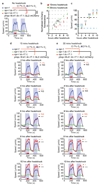
a, Bordering and aggregation phenotypes. n=4 assays. ***, p<0.001; **, p< 0.01 (ANOVA with Tukey’s correction). Data here and in all Figures show Mean ± Standard Error of the Mean. b, Cell surface expression of ILCR-1-GFP and ILCR-2-HA in HEK293T cells. c, Co-immunoprecipitation of ILCR-1 and ILCR-2. d, FLAG-ILC-17.1 binding to receptor transfected cells. e, FACS of cells transfected with the indicated vector/s and exposed to FLAG-ILC-17.1 or buffer. f, g, Average speed of animals kept 2 minutes at 7% O2 then 2 hours at 21% O2. Rescue lines are as in (a). Here and hereafter time intervals at 7% O2 are open, shading indicates 21% O2, and green and red bars on the X-axis indicate intervals used for statistics. n= 4 assays, 120 animals. *, p<0.05 **, p<0.01, and ***, p<0.001. Mann-Whitney U test. h, i, Speed at 7% and 21% O2 assayed 10 minutes (h) or 2 hours (i) after animals were transferred to assay plates. n = 3 assays, 90 animals. NS, not significant, ***, p<0.001. Mann-Whitney U test.
Figure 2
Figure 2. Anatomical focus of ILC-17.1 signaling.
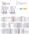
a–d ilcr-1-gfp and ilcr-2-gfp are expressed in many neurons (a, b), including RMG, identified using pflp-5::gfp (c, d). e, ilcr-1 O2 response defects rescued by selective expression in RMG (pnpr-1 and pflp-5) ***, p<0.001. Mann-Whitney U test. f, ILC-17.1 expression. Top left: Functional mCherry-ILC-17.1 accumulates in coelomocytes (labeled with GFP, ccGFP). Top right: a functional pilc-17.1::ilc-17.1::SL2::mCherry polycistronic transgene does not highlight coelomocytes but (Bottom) labels the RMG, AUA and ASE neurons. DiO-labeled neurons provide reference points. A, anterior; P, posterior. g, mCherry-ILC-17.1 accumulation in coelomocytes is not O2-dependent. NS, not significant. Student’s ṯ-test. h, Gut-specific expression of ilc-17.1 cDNA rescues ilc-17.1 defects. ***, p<0.001. Mann-Whitney U test.
Figure 3
Figure 3. ILC-17.1 signaling alters RMG physiology.
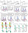
a, b, O2-evoked Ca2+ responses in URX (a) and RMG (b). NS, not significant, **, p<0.01. Mann-Whitney U test. c, Effect of stimulating RMG using Channelrhodopsin in animals kept at 7% O2. ***, p<0.001, Mann-Whitney U test. d, e, Disrupting ilc-17.1 reduces ascaroside-evoked (asc) responses in RMG (d) and switches valence of ascaroside pheromones (e). f, Tapping restores responsiveness to 21% O2 to ilc-17.1 mutants. The arrow and purple line indicate taps to the microscope stage at 7 minutes. mec-4 mutants are touch insensitive. g, h, Heat-shock induced expression in adults rescues ilc-17.1 O2 responses. Assays were performed 2 hours after animals were picked to the assay plates. The assays in (h) were done 8 hours after heat-shock. NS, not significant, ***, p<0.001 (Mann-Whitney U test).
Figure 4
Figure 4. Overexpressing ILC-17.1 heightens responsiveness to 21% O2
a, Animals overexpressing ILC-17.1 respond more strongly to 21% O2 than npr-1 control animals. **, p<0.01 (Mann-Whitney U test). b, Overexpressing ILC-17.1 in N2 animals promotes bordering and aggregation. *, p<0.05 and ****, p<0.0001 (t test). c, N2 animals overexpressing ILC-17.1 show increased responsiveness to 21% O2. **, p<0.01 (Mann-Whitney U test). d–f, ILC-17.1 overexpression increases O2-evoked Ca2+ responses in RMG in N2 (d) and npr-1 (e) animals, but does not alter O2-evoked Ca2+ responses in URX (f). NS, not significant; *, p<0.05; **, p<0.01 (Mann-Whitney U test).
Figure 5
Figure 5. ACTL-1, PIK-1/IRAK and NFKI-1 mediate ILC-17.1 signaling in RMG.
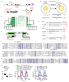
a, b, IP of ILCR-1-GFP and ILCR-2-GFP co-immunoprecipitated ACTL-1-HA (a). Conversely, IP of ACTL-1-HA pulled down ILCR-2-GFP and, more weakly, ILCR-1-GFP (b). c, d, IP of PIK-1-FLAG pulled down ACTL-1-HA (c). IP of ACTL-1-HA pulled down PIK-1-FLAG (d). e, Overexpressing NFKI-1 rescues the O2 response defects of ilcr-1, ilcr-2, actl-1 and pik-1 mutants. NS, not significant, ***, p<0.001 (Mann-Whitney U test). fh, The actl-1 (f), pik-1 (g) and nfki-1(h) O2 response defects can be rescued by selectively expressing the corresponding cDNA in RMG interneurons (pnpr-1 and pflp-5) but not URX, AQR and PQR (pgcy-32) or ASE (pflp-6) sensory neurons. *, p<0.05, ***, p<0.001 (Mann-Whitney U test). i–k, O2-evoked Ca2+ responses in RMG of single and double null mutants of ilc-17.1, actl-1, pik-1 and nfki-1. **, p<0.01, ***, p<0.001 (Mann-Whitney U test). l, ILC-17.1 signaling in C. elegans compared to IL-17 and Toll-Like Receptor (TLR) pathways in mammals. TF, transcription factor. m, Models. ILC-17.1 increases RMG responsiveness to input from the URX O2 sensors, enabling C. elegans arousal at 21% O2. Without ILC-17.1 C. elegans responds poorly to 21% O2 unless also mechanically stimulated.

Comment in

References

    1. Marder E. Neuromodulation of neuronal circuits: back to the future. Neuron. 2012;76:1–11. - PMC - PubMed
    1. Root CM, Ko KI, Jafari A, Wang JW. Presynaptic facilitation by neuropeptide signaling mediates odor-driven food search. Cell. 2011;145:133–144. - PMC - PubMed
    1. Lee SH, Dan Y. Neuromodulation of brain states. Neuron. 2012;76:209–222. - PMC - PubMed
    1. Su CY, Wang JW. Modulation of neural circuits: how stimulus context shapes innate behavior in Drosophila. Curr Opin Neurobiol. 2014;29:9–16. - PMC - PubMed
    1. Owald D, Waddell S. Olfactory learning skews mushroom body output pathways to steer behavioral choice in Drosophila. Curr Opin Neurobiol. 2015;35:178–184. - PMC - PubMed

Publication types

MeSH terms