

Dermatoscopy-guided therapy of pigmented basal cell carcinoma with imiquimod
- PMID: 28099598
- PMCID: PMC5193187
- DOI: 10.1590/abd1806-4841.20165255
Dermatoscopy-guided therapy of pigmented basal cell carcinoma with imiquimod
Abstract
Background:: Dermatoscopy is a non-invasive diagnostic tool used to examine skin lesions with an optical magnification. It has been suggested as a useful tool for monitoring therapeutic response in lentigo maligna patients treated with imiquimod.
Objective:: To examine the accuracy of dermatoscopy as a tool to monitor the therapeutic response of pigmented basal cell carcinoma treated with imiquimod.
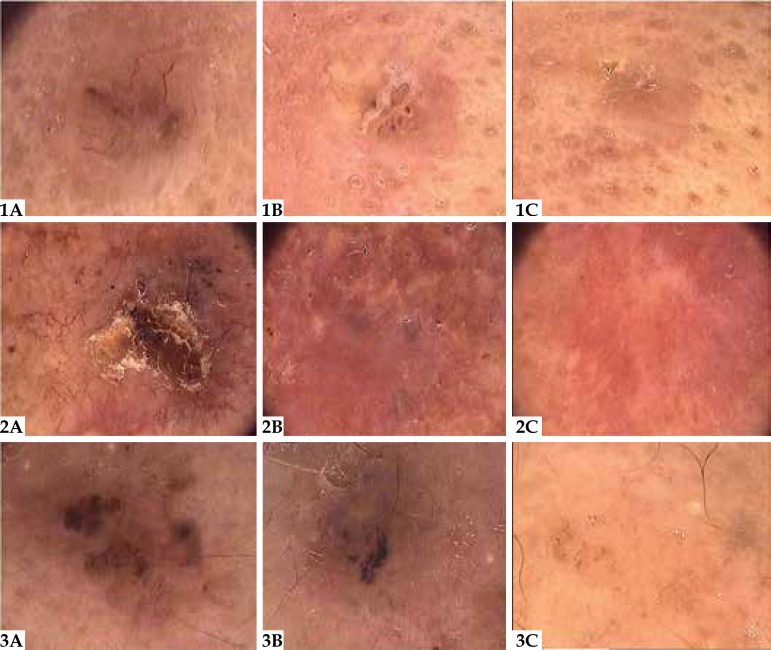
Method:: The authors designed a prospective study. Patients with pigmented basal cell carcinoma were included and data regarding the dermatoscopy features were collected following the Menzies criteria, prior to initiating the imiquimod treatment. Subsequent dermatoscopic evaluations were performed at weeks 4 and 8, following imiquimod discontinuation.
Results:: Twenty lesions were included. The most common pigmented dermatoscopy features were large blue-grey ovoid nests (80%), followed by blue-grey globules (50%) and leaf-like areas (30%). No spoke wheel areas were observed. In 17 out of 20 patients, a response was noted during the first evaluation at 4 weeks, while the clearance was noted at the second check-up after 8 weeks. In two patients, the clearance was found at the initial evaluation at 4 weeks, while in one patient, the response remained unchanged. Blue-grey globules were the fastest to exhibit clearance (50% at week 4), followed by leaf-like areas (15%) and large blue-grey ovoid nests (6.25%).
Conclusion:: According to our results, dermatoscopic evaluation enhances the accuracy in the assessment of the clinical response to imiquimod in pigmented basal cell carcinoma.
Conflict of interest statement
None
Figures
Similar articles
-
[Dermoscopic signs as predictors of non-response to imiquimod treatment in superficial basal cell carcinoma].An Sist Sanit Navar. 2019 Dec 5;42(3):303-307. doi: 10.23938/ASSN.0722. An Sist Sanit Navar. 2019. PMID: 31859267 Spanish.
-
The use of dermatoscopy to monitor therapeutic response of Bowen disease: a dermatoscopic pathological study.Br J Dermatol. 2012 Dec;167(6):1382-5. doi: 10.1111/j.1365-2133.2012.11124.x. Br J Dermatol. 2012. PMID: 22759263
-
Role of In Vivo Reflectance Confocal Microscopy in the Analysis of Melanocytic Lesions.Acta Dermatovenerol Croat. 2018 Apr;26(1):64-67. Acta Dermatovenerol Croat. 2018. PMID: 29782304 Review.
-
Lentigo maligna treated with topical imiquimod: dermatoscopy usefulness in clinical monitoring.An Bras Dermatol. 2011 Jul-Aug;86(4):792-4. doi: 10.1590/s0365-05962011000400028. An Bras Dermatol. 2011. PMID: 21987152 English, Portuguese.
-
Dermoscopic features of basal cell carcinoma and its subtypes: A systematic review.J Am Acad Dermatol. 2021 Sep;85(3):653-664. doi: 10.1016/j.jaad.2019.11.008. Epub 2019 Nov 7. J Am Acad Dermatol. 2021. PMID: 31706938 Free PMC article.
Cited by
-
Line-Field Confocal Optical Coherence Tomography May Enhance Monitoring of Superficial Basal Cell Carcinoma Treated with Imiquimod 5% Cream: A Pilot Study.Cancers (Basel). 2021 Sep 30;13(19):4913. doi: 10.3390/cancers13194913. Cancers (Basel). 2021. PMID: 34638396 Free PMC article.
-
Basal Cell Carcinoma After High Dose Rate Brachytherapy: Medium-term Dermoscopic Evaluation of Cancer's Response.Dermatol Ther (Heidelb). 2023 Sep;13(9):2063-2078. doi: 10.1007/s13555-023-00981-5. Epub 2023 Aug 9. Dermatol Ther (Heidelb). 2023. PMID: 37558829 Free PMC article.
-
Dermoscopy Update: Review of Its Extradiagnostic and Expanding Indications and Future Prospects.Dermatol Pract Concept. 2019 Oct 31;9(4):253-264. doi: 10.5826/dpc.0904a02. eCollection 2019 Oct. Dermatol Pract Concept. 2019. PMID: 31723457 Free PMC article.
-
Basal Cell Carcinoma Treated with High Dose Rate (HDR) Brachytherapy-Early Evaluation of Clinical and Dermoscopic Patterns during Irradiation.Cancers (Basel). 2021 Oct 16;13(20):5188. doi: 10.3390/cancers13205188. Cancers (Basel). 2021. PMID: 34680336 Free PMC article.
-
Dermoscopy of Basal Cell Carcinoma Part 3: Differential Diagnosis, Treatment Monitoring and Novel Technologies.Cancers (Basel). 2025 Mar 19;17(6):1025. doi: 10.3390/cancers17061025. Cancers (Basel). 2025. PMID: 40149358 Free PMC article. Review.
References
-
- Terstappen K, Larkö O, Wennberg AM. Pigmented basal cell carcinoma-comparing the diagnostic methods of SIAscopy and dermoscopy. Acta Derm Venereol. 2007;87:238–242. - PubMed
-
- Maloney ME, Jones DB, Sexton FM. Pigmented basal cell carcinoma; investigation of 70 cases. J Am Acad Dermatol. 1992;27:74–78. - PubMed
-
- Bleehen S. Pigmented basal cell epithelioma. Br J Dermatol. 1975;93:361–370. - PubMed
-
- Tezuka T, Ohkuma M, Hirose I. Melanosomes of pigmented basal cell epitheliomas. Dermatologica. 1977;154:14–22. - PubMed
-
- Krähn G, Gottlöber P, Sander C, Peter RU. Dermatoscopy and high frequency sonography two useful non-invasive methods to increase preoperative diagnostic accuracy in pigmented skin lesions. Pigment Cell Res. 1998;11:151–154. - PubMed
MeSH terms
Substances
LinkOut - more resources
Full Text Sources
Other Literature Sources
Medical