Granuloma faciale: clinical, morphological and immunohistochemical aspects in a series of 10 patients
- PMID: 28099604
- PMCID: PMC5193193
- DOI: 10.1590/abd1806-4841.20164628
Granuloma faciale: clinical, morphological and immunohistochemical aspects in a series of 10 patients
Abstract
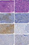
Granuloma faciale is a chronic, benign, cutaneous vasculitis with well-established clinical and morphological patterns, but with an unknown etiology. This study describes clinical and pathologic aspects of patients diagnosed with granuloma faciale. The authors analyzed demographic, clinical, morphological and immunohistochemical data from patients with a final diagnosis of granuloma faciale, confirmed between 1998 and 2012. There was a proportional and mixed inflammatory infiltrate, Grenz zones were present in almost all the samples. Immunophenotyping confirmed a higher intensity of T lymphocytes than B lymphocytes in thirteen samples, with a predominance of T CD8 lymphocytes in 64% of cases, in contrast to the literature, which indicates that the major component is T CD4 lymphocytes. All cases were positive for IgG4 but the majority (12/14) had less than 25% of stained cells. The pathogenesis of granuloma faciale remains poorly understood, making studies of morphological and immunohistochemical characterization important to better understand it.
Conflict of interest statement
None
Figures


References
-
- Rodriguez M, Richaud C. Granuloma facial a propósito de un caso. Rev Cent Dermatol Pascuan. 2001;10:147–150.
-
- Barreto AMW, Purin KSM, Fillus Neto J. Granuloma facial. An Bras Dermatol. 1991;66:25–28.
-
- Cesinaro AM, Lonardi S, Facchetti F. Granuloma Faciale - A Cutaneous Lesion Sharing Features With IgG4-associated Sclerosing Diseases. Am J Surg Pathol. 2013;37:66–73. - PubMed
-
- Ziemer M, Koehler MJ, Weyers W. Erythema elevatum diutinum - a chronic leukocytoclastic vasculitis microscopically indistinguishable from granuloma faciale. J Cutan Pathol. 2011;38:876–883. - PubMed
-
- Ortonne N, Wechsler J, Bagot M, Grosshans E, Cribier B. Granuloma faciale a clinicopathologic study of 66 patients. J Am Acad Dermatol. 2005;53:1002–1009. - PubMed
Publication types
MeSH terms
LinkOut - more resources
Full Text Sources
Other Literature Sources
Research Materials
