. 2017 Jan 17;18(3):804-815.
doi: 10.1016/j.celrep.2016.12.068.

Screening Bioactives Reveals Nanchangmycin as a Broad Spectrum Antiviral Active against Zika Virus

Affiliations

Screening Bioactives Reveals Nanchangmycin as a Broad Spectrum Antiviral Active against Zika Virus

Keiko Rausch et al. Cell Rep. .

Abstract

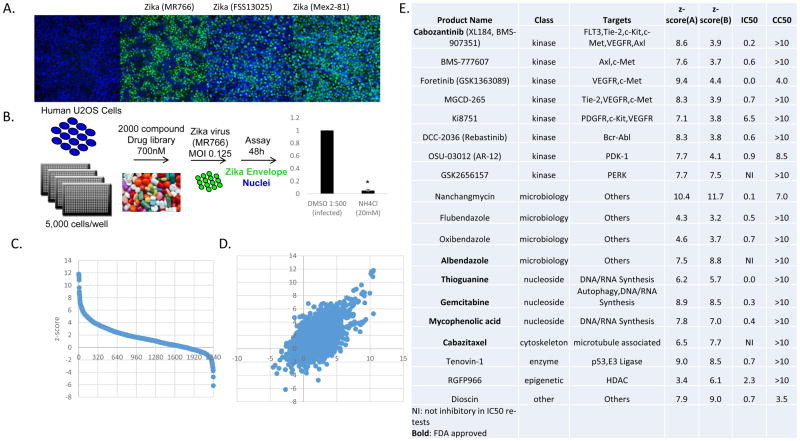
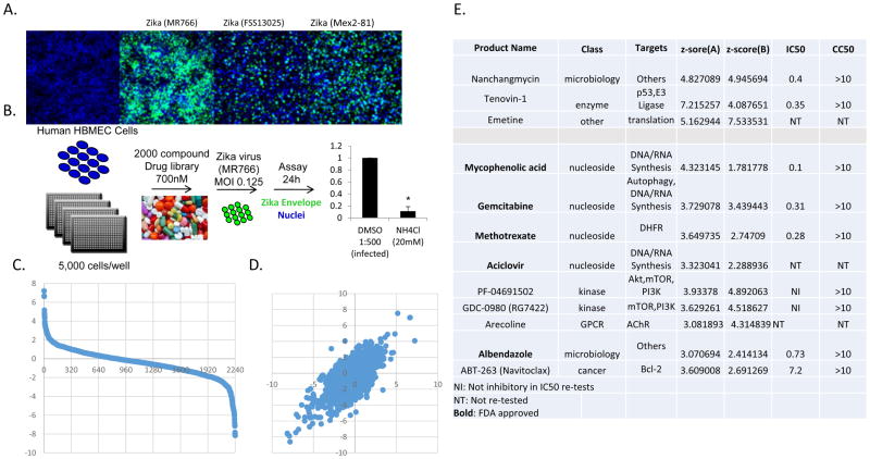
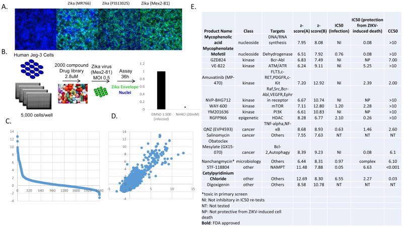
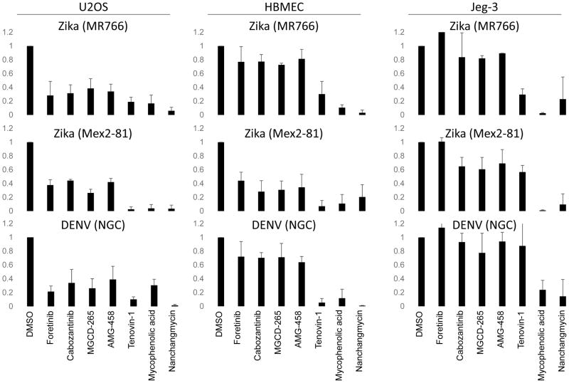
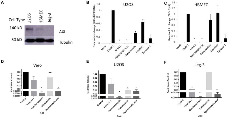
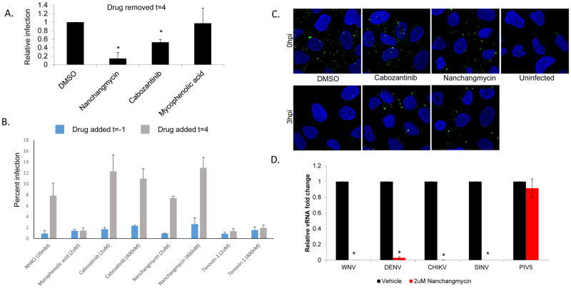
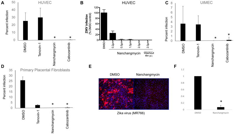
Zika virus is an emerging arthropod-borne flavivirus for which there are no vaccines or specific therapeutics. We screened a library of 2,000 bioactive compounds for their ability to block Zika virus infection in three distinct cell types with two different strains of Zika virus. Using a microscopy-based assay, we validated 38 drugs that inhibited Zika virus infection, including FDA-approved nucleoside analogs. Cells expressing high levels of the attachment factor AXL can be protected from infection with receptor tyrosine kinase inhibitors, while placental-derived cells that lack AXL expression are insensitive to this inhibition. Importantly, we identified nanchangmycin as a potent inhibitor of Zika virus entry across all cell types tested, including physiologically relevant primary cells. Nanchangmycin also was active against other medically relevant viruses, including West Nile, dengue, and chikungunya viruses that use a similar route of entry. This study provides a resource of small molecules to study Zika virus pathogenesis.

Keywords: antivirals; arbovirus; drugs; entry; flavivirus; repurposing; therapeutics.


Figures

Figure 1
Figure 1
19 drugs robustly block ZIKV infection in human U2OS cells. (A) U2OS cells were infected with three different strains of ZIKA virus and the infection was monitored by microscopy (anti-flavivirus envelope, 4G2, green; nuclei, blue). (B) Schematic of the screening strategy with ZIKV (MR766) where ammonium chloride (NH4Cl) was used as a positive control. (C) Z scores of the percent infection of replicate 1. (D) Z scores of replicate 1 plotted against replicate 2. (E) The 19 drugs that inhibit infection with Z-scores>3, impacted infection >3-fold that are nontoxic. IC50 values for ZIKV (Mex2-81) (uM) and CC50 values (uM) shown.
Figure 2
Figure 2
12 drugs robustly block ZIKV infection in human HBMEC cells. (A) HBMEC cells were infected with three different strains of ZIKV virus and the infection was monitored by microscopy (anti-flavivirus envelope, 4G2, green; nuclei, blue). (B) Schematic of the screening strategy where ammonium chloride (NH4Cl) was used as a positive control. (C) Z-scores of the percent infection of replicate 1. (D) Z-scores of replicate 1 plotted against replicate 2. (E) The 12 drugs that inhibit infection >3-fold that are nontoxic. IC50 values for ZIKV (Mex2-81) (uM) and CC50s are shown
Figure 3
Figure 3
16 drugs robustly block ZIKV infection in human Jeg-3 cells. (A) Jeg-3 cells were infected with three different strains of ZIKV and the infection was monitored by microscopy (anti-flavivirus envelope, 4G2, green; nuclei, blue). (B) Schematic of the screening strategy where ammonium chloride (NH4Cl) was used as a positive control. (C) Z-scores of the percent infection of replicate 1. (D) Z-scores of replicate 1 plotted against replicate 2. (E) The 15 drugs that inhibit infection >3-fold that are nontoxic. IC50 values for ZIKV (Mex2-81) (uM), IC50s for protection from ZIKV-induced death for ZIKV (Mex2-81) (uM), and CC50 values are shown.
Figure 4
Figure 4
Anti-ZIKV inhibitors show activity against DENV. The indicated cell lines were treated with the indicated drugs at 2uM one hour prior to infection with the indicated flaviviruses and the percent infection was quantified using automated microscopy from 3 independent experiments with mean±SD of the fold change in infection shown.
Figure 5
Figure 5
Receptor tyrosine kinase inhibitors block infection only in cells that express high levels of the entry factor AXL. (A) The indicated cell lines were processed for immunoblot for AXL and the control tubulin. A representative experiment is shown. U2OS (B) or HBMEC (C) cells were treated with the indicated drugs and infected with ZIKV (Mex2-81, MOI=2.5) and processed for RT-qPCR analysis to monitor the level of infection from 3 independent experiments with mean±SD shown. (D–F) The indicated cell type was treated with the indicated drugs and infected with ZIKV (Mex2-81) and TCID50s were calculated for 3 independent experiments with mean±SD shown. *p,0.05.
Figure 6
Figure 6
Nanchangmycin potently blocks infection at the level of entry. (A) U2OS cells were treated with the indicated drugs and infected with ZIKV (Mex2-81, MOI 25), 4 hpi the drugs were washed out and the cells were processed for immunofluorescence 24hpi from 3 independent experiments with mean±SD shown. (B) U2OS cells were treated with the indicated drugs either one hour prior to infection (blue) or 4hpi (grey) with ZIKV (Mex2-81, MOI=1). NH4Cl was added at 4hpi to block further entry. The infection was processed at 24hpi for microscopy and the percent infection shown for a representative experiment of three. (C) U2OS cells were treated with the indicated drugs and then infected with ZIKV (Mex2-81, MOI=100) at 4C for one hour for binding or released at 37C for 3h to allow uptake. The cells were fixed without permeablization to monitor extracellular virions (anti-envelope, green; nuclei, blue). Images were taken by confocal microscopy and are representative of 3 independent experiments. (D) U2OS cells were treated with vehicle or nanchangmycin (2uM) and infected with the indicated viruses for 24h and processed for RT-qPCR with mean±SD shown. * p<0.05.
Figure 7
Figure 7
Nanchangmycin blocks ZIKV infection in primary cells. (A–B) HUVEC, (C) UtMEC, (D) or primary placental fibroblasts were treated with the indicated drugs and infected by ZIKV (FSS13025, MOI=1) and processed for immunofluorescence at 48 hpi. The percentage of infected cells was calculated and mean±SD shown; *p<0.05. (E–F) Murine mixed neuronal cultures were infected with ZIKA (MR766, MOI=2) for 24hr and processed for immunofluorescence. (E) Representative images shown for 3 independent experiments quantified in (F) with mean±SD shown; *p<0.05.

References

    1. Araujo AQ, Silva MT, Araujo AP. Zika virus-associated neurological disorders: a review. Brain. 2016;139:2122–2130. - PubMed
    1. Barrows NJ, Campos RK, Powell ST, Prasanth KR, Schott-Lerner G, Soto-Acosta R, Galarza-Munoz G, McGrath EL, Urrabaz-Garza R, Gao J, et al. A Screen of FDA-Approved Drugs for Inhibitors of Zika Virus Infection. Cell Host Microbe. 2016;20:259–270. - PMC - PubMed
    1. Barzon L, Trevisan M, Sinigaglia A, Lavezzo E, Palu G. Zika virus: From pathogenesis to disease control. FEMS Microbiol Lett 2016 - PubMed
    1. Bayer A, Lennemann NJ, Ouyang Y, Bramley JC, Morosky S, Marques ET, Jr, Cherry S, Sadovsky Y, Coyne CB. Type III Interferons Produced by Human Placental Trophoblasts Confer Protection against Zika Virus Infection. Cell Host Microbe. 2016;19:705–712. - PMC - PubMed
    1. Blitvich BJ, Firth AE. Insect-specific flaviviruses: a systematic review of their discovery, host range, mode of transmission, superinfection exclusion potential and genomic organization. Viruses. 2015;7:1927–1959. - PMC - PubMed

MeSH terms