

Gestational diabetes mellitus and retinal microvasculature
- PMID: 28100181
- PMCID: PMC5241913
- DOI: 10.1186/s12886-016-0398-7
Gestational diabetes mellitus and retinal microvasculature
Abstract
Background: Small-vessel dysfunction may be an important consequence of chronic hyperglycemia. We examined the association between gestational diabetes mellitus (GDM), a state of transient hyperglycemia during pregnancy, and retinal microvascular changes in pregnant women at 26-28 weeks of pregnancy.
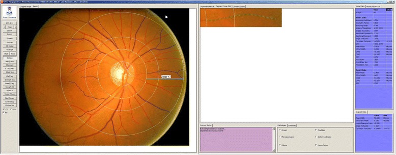
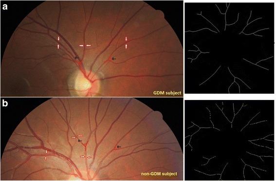
Methods: A total of 1136 pregnant women with singleton pregnancies were recruited during their first trimester at two major Singapore maternity hospitals in an on-going birth cohort study. Participants underwent an oral glucose tolerance test and retinal imaging at 26-28 weeks gestation (n = 542). We used the 1999 World Health Organization (WHO) criteria to define GDM: ≥7.0 mmol/L for fasting glucose and/or ≥7.8 mmol/L for 2-h post-glucose. Retinal microvasculature was measured using computer software (Singapore I Vessel Analyzer, SIVA version 3.0, Singapore Eye Research Institute, Singapore) from the retinal photographs.
Results: In a multiple linear regression model adjusting for age, ethnicity and maternal education, mothers with GDM had narrower arteriolar caliber (-1.6 μm; 95% Confidence Interval [CI]: -3.1 μm, -0.2 μm), reduced arteriolar fractal dimension (-0.01 Df; 95% CI: -0.02 Df, -0.001 Df;), and larger arteriolar branching angle (1.8°; 95% CI: 0.3°, 3.3°) than mothers without GDM. After further adjusting for traditional risks of GDM, arteriolar branching angle remained significantly larger in mothers with GDM than those without GDM (2.0°; 95% CI: 0.5°, 3.6°).
Conclusions: GDM was associated with a series of retinal arteriolar abnormalities, including narrower caliber, reduced fractal dimension and larger branching angle, suggesting that transient hyperglycemia during pregnancy may cause small-vessel dysfunction.
Keywords: Gestational diabetes mellitus; Pregnancy outcomes; Retinal imaging; Retinal microvascular measures; Retinal microvasculature.
Figures
References
MeSH terms
LinkOut - more resources
Full Text Sources
Other Literature Sources
Miscellaneous
