. 2017 Mar 1;312(3):R443-R450.
doi: 10.1152/ajpregu.00270.2016. Epub 2017 Jan 18.

Regional diaphragm volume displacement is heterogeneous in dogs

Affiliations

Regional diaphragm volume displacement is heterogeneous in dogs

Brooke Greybeck et al. Am J Physiol Regul Integr Comp Physiol. .

Abstract

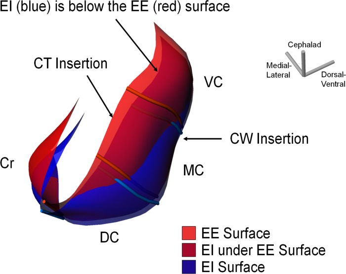
Muscle shortening and volume displacement (VD) are critical determinants of the pressure-generating capacity of the diaphragm. The present study was designed to test the hypothesis that diaphragm VD is heterogeneous and that distribution of VD is dependent on regional muscle shortening, posture, and the level of muscle activation. Radioopaque markers were sutured along muscle bundles of the peritoneal surface of the crural, dorsal costal, midcostal, and ventral costal regions of the left hemidiaphragm in four dogs. The markers were followed by biplanar video fluoroscopy during quiet spontaneous breathing, passive inflation to total lung capacity (TLC), and inspiratory efforts against an occluded airway at three lung volumes spanning the vital capacity [functional residual capacity, functional residual capacity + ½ inspiratory capacity, and TLC in both the prone and supine postures]. Our data show the ventral costal diaphragm had the largest VD and contributed nearly two times to the total diaphragm VD compared with the dorsal costal portion. In addition, the ventral costal diaphragm contributed nearly half of the total VD in the prone position, whereas it only contributed a quarter of the total VD in the supine postition. During efforts against an occluded airway and during passive inflation to TLC in the supine position, the crural diaphragm displaced volume equivalent to that of the midcostal portion. Regional muscle shortening closely matched regional VD. We conclude that the primary force generator of the diaphragm is primarily dominated by the contribution of the ventral costal region to its VD.

Keywords: chest wall mechanics; modeling diaphragm kinematics; respiratory muscle.


Figures

Fig. 1.
Fig. 1.
Surface of the left hemidiaphragm at the end of expiration (EE) during quiet spontaneous breathing in a representative prone dog. The position of muscle fibers in each of the four regions [ventral costal (VC), midcostal (MC), dorsal costal (DC), and crural (Cr)] is shown. Nine muscle fibers (brown), excluding the Cr-costal boundary, were used to create surfaces to study volume displacement (VD) in the costal and Cr diaphragm. The central tendon (CT) insertion and chest wall (CW) insertion are also indicated on the diagram.
Fig. 2.
Fig. 2.
Two surfaces of the diaphragm at the EE (red) and end of inspiration (EI; blue) during spontaneous quiet breathing in the same prone dog shown in Fig. 1. The surfaces were divided into the four regions of the diaphragm (VC, MC, DC, and Cr) by the muscle fibers shown. The diaphragm at the EI descends along the CT insertion site as it contracts, which is shown as being underneath the surface at the EE. This is shown in the dark red color.
Fig. 3.
Fig. 3.
Abdominal (green) and pleural (pink) volumes displaced by the diaphragm during spontaneous breathing in the same prone dog as shown in Figs. 1 and 2. The surfaces of the diaphragm at the EE (red) and EI (blue) cross along a line of intersection, thereby displacing two distinct volumes: the abdominal and pleural volumes. Note that the dorsal region of the costal diaphragm displaced the most pleural VD compared with the other regions of the diaphragm.
Fig. 4.
Fig. 4.
Percent total volume displaced (VD) (abdominal VD + pleural VD) by the four regions of the diaphragm (VC, MC, DC, and Cr) as a function of posture (prone and supine). Total VD was determined for the each of the modes of ventilation [spontaneous quiet breathing, occluded (Occ) at functional residual capacity (FRC), occluded at functional residual capacity plus half inspiratory capacity (FRC + ½IC), occluded at total lung capacity (TLC), and passive inflation to total lung capacity (passive TLC)]. While regional changes in total VD stayed consistent throughout changes in posture and mode of ventilation, there were apparent regional variations within each posture and ventilation state (P < 0.001).
Fig. 5.
Fig. 5.
Percent net volume displaced (VD) (abdominal VD – pleural VD) by the four regions of the diaphragm (VC, MC, DC, and Cr) as a function of posture [prone (A) and supine (B)] and mode of ventilation (spontaneous quiet breathing, occluded at FRC, occluded at FRC + ½IC, occluded at TLC, and passive inflation to TLC). Error bars on both graphs represent SEs. There was a large contribution of pleural VD compared with abdominal VD in the DC portion of the diaphragm, as indicated by the negative net VD in both the prone and supine postures. During spontaneous breathing, there was significantly more contribution of the pleural VD in the prone posture compared with the supine posture (P < 0.001). There was a significant variation in volume displaced by each region of the diaphragm in both postures (P < 0.001). As lung volume increased against the occluded airway, dogs under the prone position exhibited a decrease in pleural VD in the DC region of the diaphragm. No significant changes in net volume displaced were observed as a function of mode of ventilation in the supine posture.
Fig. 6.
Fig. 6.
Percent muscle shortening within each of the four regions (VC, MC, DC, and Cr) at two different postures [supine (A) and prone (B)]. Average percent muscle shortening was computed for all breathing maneuvers: spontaneous quiet breathing, occluded at FRC, occluded at FRC + ½IC, occluded at TLC, and passive inflation to TLC. SEs are represented by the bars in each graph. ANOVA showed a significant decrease between the percent muscle shortening during spontaneous breathing compared with all other ventilatory maneuvers in all regions of the costal and Cr diaphgram for both postures (*P < 0.01). There were no significant differences in percent muscle shortening between the regions for all occluded efforts; however, there was a significant reduction of the percent muscle shortening in the dorsal region during spontaneous breathing compared with the percent muscle shortening in the other regions of the diaphragm during spontaneous breathing (#P = 0.04).
Fig. 7.
Fig. 7.
Percent muscle shortening of the different regions of the costal diaphragm (refer to inset schematic for region identification) compared with the regional blood flow per gram of muscle. Blood flow was normalized as a ratio of the regional blood flow to that measured in costal region 7 during rest, as determined by Johnson et al. (19). Percent muscle shortening in the costal diaphragm coincided with the normalized regional blood flow, with increasing muscle shortening and blood flow occurring from ventral to MC regions and then decreasing muscle shortening and blood flow from MC to dorsal regions.

References

    1. Amancharla MR, Rodarte JR, Boriek AM. Modeling the kinematics of the canine midcostal diaphragm. Am J Physiol Regul Integr Comp Physiol 280: R588–R597, 2001. - PubMed
    1. Angelillo M, Boriek AM, Rodarte JR, Wilson TA. Shape of the canine diaphragm. J Appl Physiol 89: 15–20, 2000. - PubMed
    1. Angelillo M, Boriek AM, Rodarte JR, Wilson TA. Theory of diaphragm structure and shape. J Appl Physiol 83: 1486–1491, 1997. - PubMed
    1. Boriek AM, Black B, Hubmayr R, Wilson TA. Length and curvature of the dog diaphragm. J Appl Physiol 101: 794–798, 2006. doi:10.1152/japplphysiol.00865.2004. - DOI - PubMed
    1. Boriek AM, Liu S, Rodarte JR. Costal diaphragm curvature in the dog. J Appl Physiol 75: 527–533, 1993. - PubMed

Publication types

LinkOut - more resources