. 2017 Mar;205(3):1229-1245.
doi: 10.1534/genetics.116.197343. Epub 2017 Jan 18.

Axon Termination, Pruning, and Synaptogenesis in the Giant Fiber System of Drosophila melanogaster Is Promoted by Highwire

Affiliations

Axon Termination, Pruning, and Synaptogenesis in the Giant Fiber System of Drosophila melanogaster Is Promoted by Highwire

Melissa Borgen et al. Genetics. 2017 Mar.

Abstract

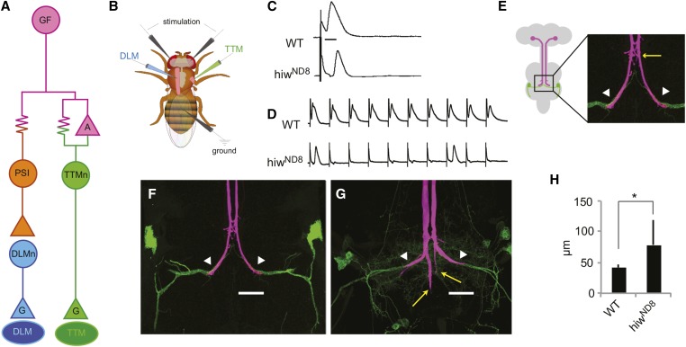
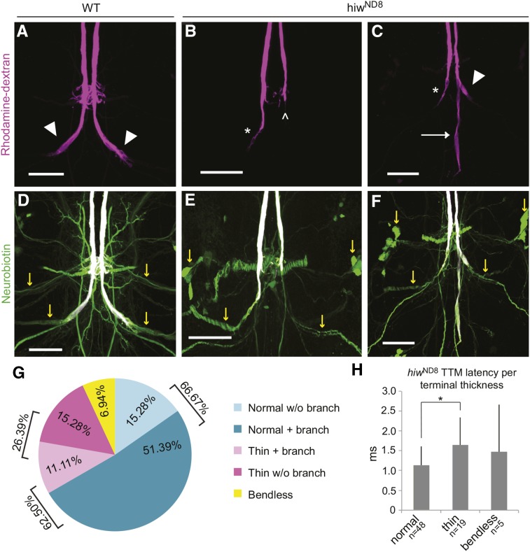
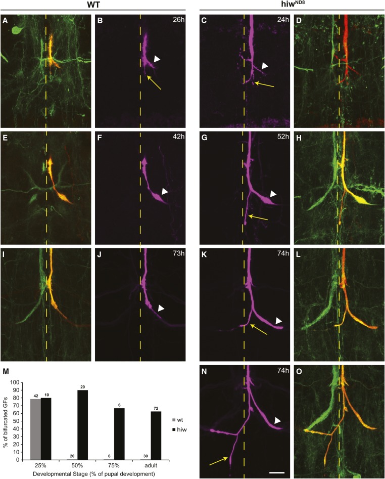
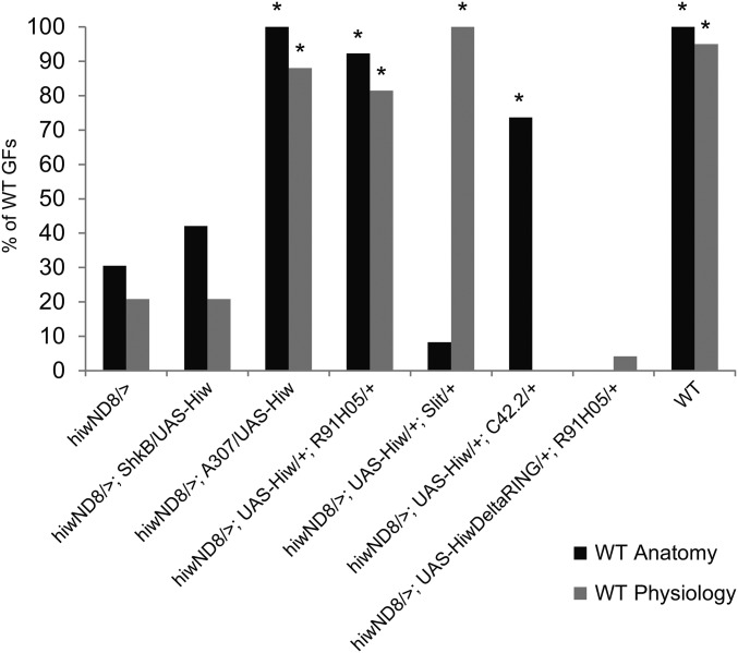
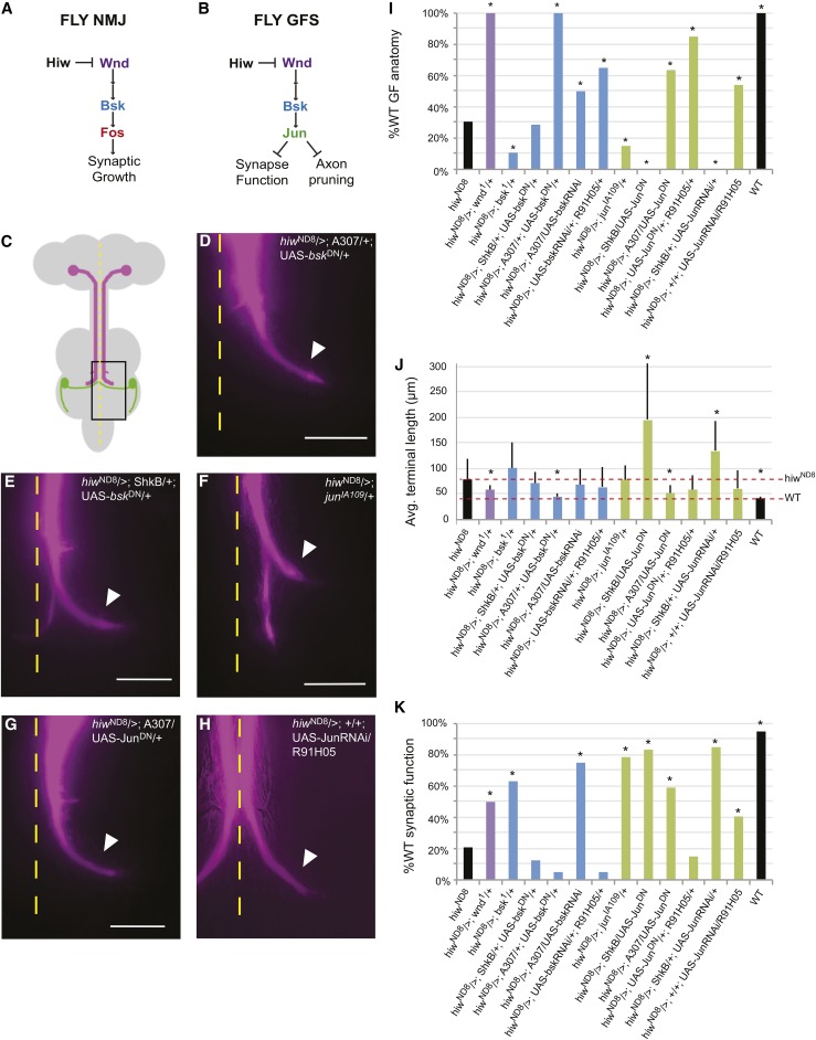
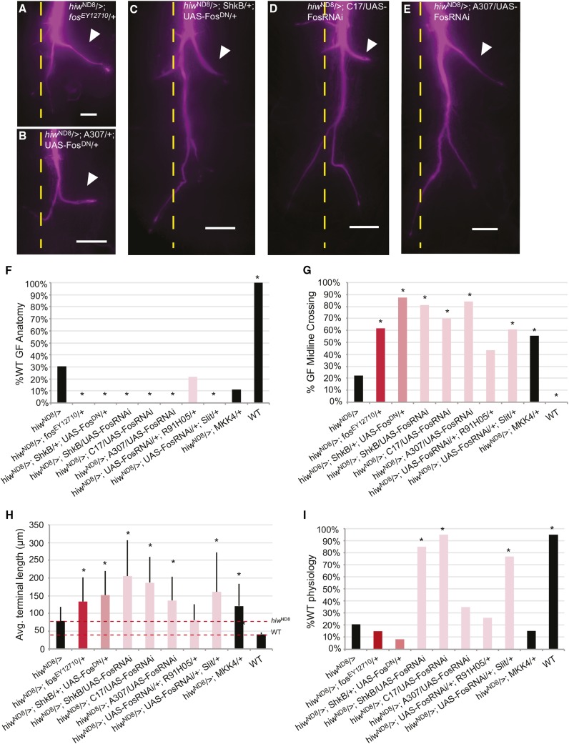
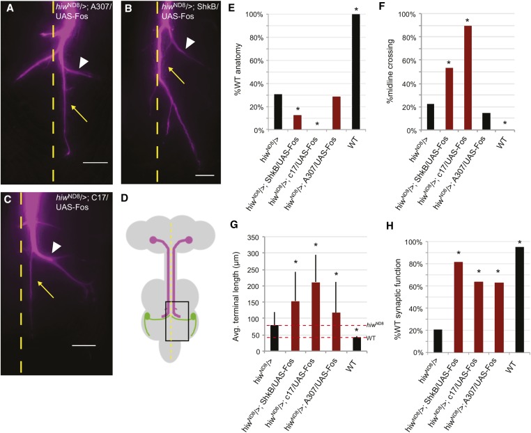
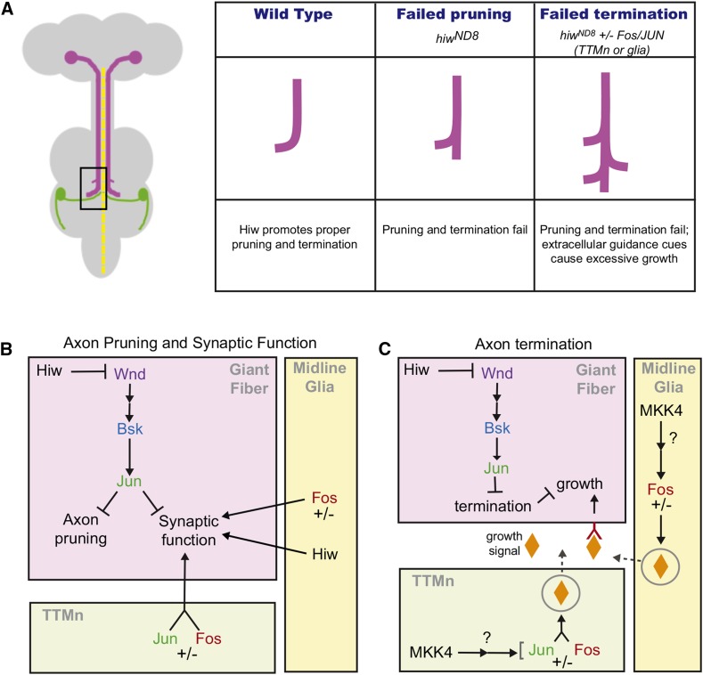
The ubiquitin ligase Highwire has a conserved role in synapse formation. Here, we show that Highwire coordinates several facets of central synapse formation in the Drosophila melanogaster giant fiber system, including axon termination, axon pruning, and synaptic function. Despite the similarities to the fly neuromuscular junction, the role of Highwire and the underlying signaling pathways are distinct in the fly's giant fiber system. During development, branching of the giant fiber presynaptic terminal occurs and, normally, the transient branches are pruned away. However, in highwire mutants these ectopic branches persist, indicating that Highwire promotes axon pruning. highwire mutants also exhibit defects in synaptic function. Highwire promotes axon pruning and synaptic function cell-autonomously by attenuating a mitogen-activated protein kinase pathway including Wallenda, c-Jun N-terminal kinase/Basket, and the transcription factor Jun. We also show a novel role for Highwire in non-cell autonomous promotion of synaptic function from the midline glia. Highwire also regulates axon termination in the giant fibers, as highwire mutant axons exhibit severe overgrowth beyond the pruning defect. This excessive axon growth is increased by manipulating Fos expression in the cells surrounding the giant fiber terminal, suggesting that Fos regulates a trans-synaptic signal that promotes giant fiber axon growth.

Keywords: Fos; giant fiber; highwire; pruning; synaptogenesis.


Figures

Figure 1
Figure 1
Highwire in the giant fiber system. (A) Circuit diagram of the GFS. The GF (magenta) commands two motor outputs. The GF forms both gap junctions (resistor symbol) and cholinergic synapses (magenta triangle “A”) with the TTMn (green), which innervates the TTM “jump” muscle (dark green oval) with a glutamatergic synapse (triangle “G”). For the DLM flight pathway, the GF electrically synapses with the PSI (orange), an interneuron that synapses cholinergically with the DLMn (blue), which innervates the DLM “flight” muscle (dark blue oval). (B) Schematic of electrophysiology experiment. Extracellular stimulating electrodes are placed in the brain, recording electrodes in the TTM and DLM muscles, and a ground in the abdomen. The location of the CNS is shown in gray. (C) Recordings from the TTM illustrate the response latencies of TTM to brain stimulation. Calibration bar = 2 msec. Top trace shows a WT response latency of ∼0.8 msec. Bottom trace shows hiwND8 response latency is 1.69 msec, about double that of WT. (D) Single sweeps illustrate response to repetitive stimulation at 100 Hz. Top trace is WT and each stimulus is followed by a response. Bottom trace shows hiwND8, which does not follow high-frequency stimulation 1:1. In this case, the TTM only responded after the first and eighth stimuli. (E) Anatomy of the GF system. Schematic representation of the fly CNS, showing location of GFs (magenta) and TTMn (green). The presynaptic terminal in the second thoracic neuromere (box) is shown as a confocal image. The site of the GF:PSI synapse (yellow arrow) is used as the starting point to measure the length of the axon terminal and the white arrowhead as the end of the terminal. (F) WT GF axon terminals (magenta and white arrowheads) synapsing with TTMn medial dendrites (green). The TTMn somata are shown at the edge of the frame. (G) Mutant hiwND8 GF axons synapse with TTMn, but project ectopic branches caudally past the target (yellow arrows). (H) Average terminal length of WT and hiwND8 mutants. The average length of hiwND8 terminal measured as in E is nearly double the length of the WT terminal, due to the ectopic branching. DLM, dorsal longitudinal flight muscle; DLMn, DLM neuron; GF, giant fiber; GFS, GF system; TTM, tergotrochanteral jump muscle; TTMn, TTM neuron; PSI, Peripherally Synapsing Interneuron; WT, wild-type.
Figure 2
Figure 2
Anatomical and physiological hiwND8 phenotypes. (A–C) Rhodamine dextran-filled GF terminals. (A) WT Urbana-S shows terminals of normal thickness (arrowheads). (B and C) The characteristic hiwND8 mutant phenotype exhibits caudal branches (C, arrow). Some specimens occasionally exhibit “bendless” terminals (B, carrot) and terminals that are thinner than normal (B and C, asterisks). (D–F) Neurobiotin/Rhodamine dextran colabeling of the same preparations used in A–C. Neurobiotin (green) shows colabeling with Rhodamine dextran (white) in the GFs. In D, Urbana-S shows normal dye coupling, as neurobiotin (green) fills TTMn and PSI neurons (yellow arrows). Despite morphological and physiological defects, hiwND8 mutant GFs dye couple to TTMn and PSI (E and F, yellow arrows), demonstrating that synaptic function defects are not due to missing gap junctions. (G) Distribution of male hiwND8 axon terminal thickness per phenotype (n = 72). WT refers to GFs without the caudal branch. “Branch” refers to caudal branch. “Normal,” “thin,” and “bendless” refer to the thickness of the terminal. 26.39% of hiwND8 terminals are thin. The presence of the caudal branch is not correlated with the thin terminal phenotype; 62.5% (n = 45) of males exhibit the caudal branch and only 17.78% (n = 8) of those males have thin terminals. Terminal thickness is normal in 66.67% of hiwND8 male GFs. Thus, we focus on the caudal branch as the strong anatomical phenotype of hiwND8. (H) Average TTM response latencies of normal, bendless, and thin GFs of hiwND8 males (n = 72). While all have longer than WT latencies, thin GFs (26.39% of terminals) have a significantly longer average TTM response latency compared to the normal sized terminals (66.67% of terminals). This suggests impedance mismatch as a possible cause of severe latency defects. GF, giant fiber; TTM, tergotrochanteral jump muscle; TTMn, TTM neuron; PSI, Peripherally Synapsing Interneuron; WT, wild-type.
Figure 3
Figure 3
Axon growth and refinement during PD. Left two columns illustrate WT terminals at various stages of development. (A and B) At ∼25% of development, the axon has reached the target area, appears to be in contact with the TTMn dendrite (white arrow head), but also exhibits extra branches projecting posteriorly (yellow arrow). (E and F) By 45% of development (I and J), the excess branching has been eliminated and the presynaptic terminal is readily identified (arrowhead). At ∼75% of PD, the terminals appear very similar to the adult terminal (arrowhead). The two right columns illustrate Hiw terminals at various stages of development. (C and D) At 25% of development, the terminals exhibit excess branching and are very similar to the wild-type terminals. (G and H) At ∼45% of PD, the lack of pruning is clear in the hiwND8 and the mutant extension is obvious (yellow arrow). (K and L, and N and O) at ∼75% of PD the excess branching (arrow) is still present. These two examples illustrate the variability because the left GF (labeled by GFP) appears normal, but the dye-injected right GF exhibits excess branching as well as crossing the midline (yellow arrows). (M) Overgrowth and retraction of the GF terminal in control and mutant animals. Note the excess branching is at the peak in WT at ∼25% and most branches are retracted by 50%. The excess branching persists throughout development in the mutant. All animals were raised at 25°; at this temperature, PD occurs in 100 hr, making hours and % of PD interchangeable. GF, giant fiber; PD, pupal development; WT, wild-type.
Figure 4
Figure 4
The rescue of hiwND8 phenotypes by tissue-specific expression of UAS-Hiw. The presynaptic expression of UAS-Hiw by the R91H05 driver significantly rescued both structure and function. Postsynaptic expression with the ShkB driver did not rescue either structure or function. Midline glial expression (Slit driver) rescued synaptic function, but not axon structure, revealing a non-cell autonomous role for Hiw in synaptic function. Expression of UAS-Hiw late in PD (C42.2 driver) rescued axon morphology, but not synaptic function. Presynaptic expression of a Hiw missing its RING domain with the R91H05 driver failed to rescue morphology or synaptic function, showing that the ligase activity of Hiw is required. PD, pupal development; RING, really interesting new gene; WT, wild-type.
Figure 5
Figure 5
Knocking down the MAPK pathway rescued the hiwND8 phenotype. (A) The MAPK pathway as proposed for the fly NMJ. (B) The MAPK pathway proposed for the fly GFS. (C) Cartoon representation of the fly CNS. Box denotes area shown in micrographs. (D–H) Fluorescence Extended Depth of Focus images of GF axon terminals filled with Lucifer yellow (recolored magenta). ImageJ Background Subtraction was used. Calibration bar = 20 µm. Arrowhead marks normal terminal bend. The hiw mutant phenotype was rescued by the strong presynaptic knockdown of Bsk in hiwND8/>; A307/+; UAS-bskDN/+ (see D). Postsynaptic expression of the basket dominant negative hiwND8/>; ShkB/+; UAS-bskDN/+ mutants exhibits the caudal branch, showing the absence of a postsynaptic role for Bsk (see E). Mutant anatomy was not rescued by a heterozygous version of jun in a hiw background (hiwND8/>; junIA109/+) (see F). hiwND8/>; A307/UAS-JunDN provided a strong knockdown of Jun and rescued anatomy (see G). Expression of JunRNAi in the GFs hiwND8/>; +/+; UAS-JunRNAi/R91H05 also rescued anatomy (see H). (I) % WT GF anatomy, * P = ≤ 0.05 (Fisher’s exact test vs. hiwND8). (J) Average terminal lengths (micrometer), * P ≤ 0.05 (two-tailed Student’s t-test vs. hiwND8). (K) % WT GF physiology, * P ≤ 0.05 (Fisher’s exact test vs. hiwND8). Avg., average; GF, giant fiber; GFS, GF system; MAPK, mitogen-activated protein kinase; NMJ, neuromuscular junction; RNAi, RNA interference; WT, wild-type.
Figure 6
Figure 6
Knockdown of Fos in a hiwND8 background disrupts pruning. (A–E) Single GF axon terminals were filled with Lucifer yellow (recolored magenta) and images shown as Extended Depth of Focus compressed stacks. (A) hiwND8/>; fosEY12710/+ has increased terminal length compared to hiwND8. (B) hiwND8/>; A307/+; UAS-FosDN/+ axon terminal lengths were not significantly longer than hiwND8. Disruption of Fos in TTMn and midline glia significantly increased axon terminal lengths: (C) hiwND8/>; ShkB/+; UAS-FosDN/+, (D) hiwND8/>; C17/UAS-FosRNAi, and (E) hiwND8/>; A307/UAS-FosRNAi. Calibration bar = 20 µm. The GF terminal bend along the TTMn dendrite is marked by an arrowhead. (F) Percent anatomically WT GFs observed in various genotypes. Knocking down Fos non-cell autonomously in glia or TTMn enhanced GF anatomical defects. * P < 0.05 (Fisher’s exact test). (G) Percent GFS crossing the midline observed in various genotypes. Knocking down Fos strongly in the midline glia and TTMn increased midline crossing. * P < 0.05 (Fisher’s exact test). (H) Average terminal lengths (micrometer) observed in various genotypes. * P < 0.05 (two-tailed Student’s t-test). (I) Percent of GFs with WT synaptic function observed in various genotypes. Knocking down Fos strongly in the midline glia and TTMn rescued synaptic function. All mutants were compared to hiwND8. * P < 0.05 (Fisher’s exact test). Avg., average; GF, giant fiber; GFS, GF system; TTMn, tergotrochanteral jump muscle neuron; RNAi, RNA interference; WT, wild-type.
Figure 7
Figure 7
Fos overexpression in a hiwND8 background. Extended Depth of Focus compressed stacks of Lucifer yellow-filled GF terminals (recolored magenta). Calibration bar = 20 µm. Overexpression of Fos in a hiwND8 background rescued synaptic function and increased axon growth: (A) hiwND8/>; A307/UAS-Fos, (B) hiwND8/>; ShkB/UAS-Fos, (C) hiwND8/>; C17/UAS-Fos, and (D) Schematic of the CNS. Boxed area shows giant synapse region shown in A–C. (E) Percent WT GF anatomy observed in different genotypes. (F) Percent GFs that cross the midline observed in different genotypes. (G) Average terminal lengths (micrometer) observed in different genotypes. (H) Percent WT GF physiology observed in different genotypes. Mutants were compared to hiwND8. * P < 0.05; (E–G) used the Fisher’s exact test. (H) used a two-tailed Student’s t-test. Avg., average; GF, giant fiber; WT, wild-type.
Figure 8
Figure 8
Model of Hiw and MAPK activity in the GFS. (A) color coded schematic of the CNS with various giant fiber phenotypes schematized. (B) Hiw acts in the GFs to negatively regulate pruning and function via the Wnd MAPK cascade, which includes Bsk and Jun. The MAPK cascade suppresses both synaptic function and axon pruning. Names of proteins are displayed in the colors that were used in Figure 5 and Figure 6: Wnd (purple), Bsk (blue), Jun (green), and Fos (red). Hiw also acts in the glia to promote synaptic function. (C) Hiw also acts in the GFs to regulate axon termination, likely by regulating a receptor that binds a secreted growth signal(s) (orange diamond), which comes from the TTMn and/or glia; this trans-synaptic signal is controlled by Fos. Because mkk4 LOF phenocopies fos LOF in the hiwND8 background, we suspect Mkk4 may act upstream of Fos as part of a parallel MAPK canonical positive regulation pathway. GF, giant fiber; GFS, GF system; LOF, loss-of-function; MAPK, mitogen-activated protein kinase; TTMn, tergotrochanteral jump muscle neuron.

References

    1. Abrams B., Grill B., Huang X., Jin Y., 2008. Cellular and molecular determinants targeting the Caenorhabditis elegans PHR protein RPM-1 to perisynaptic regions. Dev. Dyn. 237: 630–639. - PMC - PubMed
    1. Allen M. J., Godenschwege T. A., 2010. Electrophysiological recordings from the Drosophila giant fiber system (GFS). Cold Spring Harb. Protoc. 2010: pdb prot5453. - PMC - PubMed
    1. Allen M. J., Drummond J. A., Moffat K. G., 1998. Development of the giant fiber neuron of Drosophila melanogaster. J. Comp. Neurol. 397: 519–531. - PubMed
    1. Atwood H. L., Govind C. K., Wu C. F., 1993. Differential ultrastructure of synaptic terminals on ventral longitudinal abdominal muscles in Drosophila larvae. J. Neurobiol. 24: 1008–1024. - PubMed
    1. Augustin H., Allen M. J., Partridge L., 2011. Electrophysiological recordings from the giant fiber pathway of D. melanogaster. J. Vis. Exp. 47: 2412. - PMC - PubMed

MeSH terms

LinkOut - more resources