. 2017 Mar 13;91(7):e02189-16.
doi: 10.1128/JVI.02189-16. Print 2017 Apr 1.

Dynamic Modulation of Expression of Lentiviral Restriction Factors in Primary CD4+ T Cells following Simian Immunodeficiency Virus Infection

Affiliations

Dynamic Modulation of Expression of Lentiviral Restriction Factors in Primary CD4+ T Cells following Simian Immunodeficiency Virus Infection

Andrew R Rahmberg et al. J Virol. .

Abstract

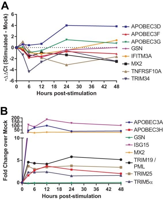
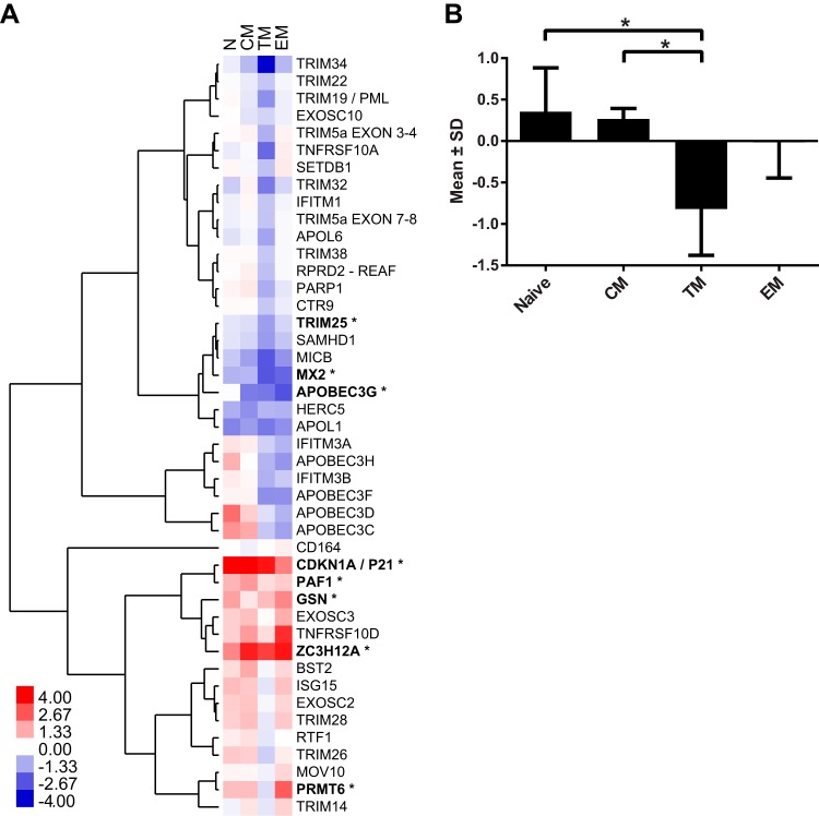
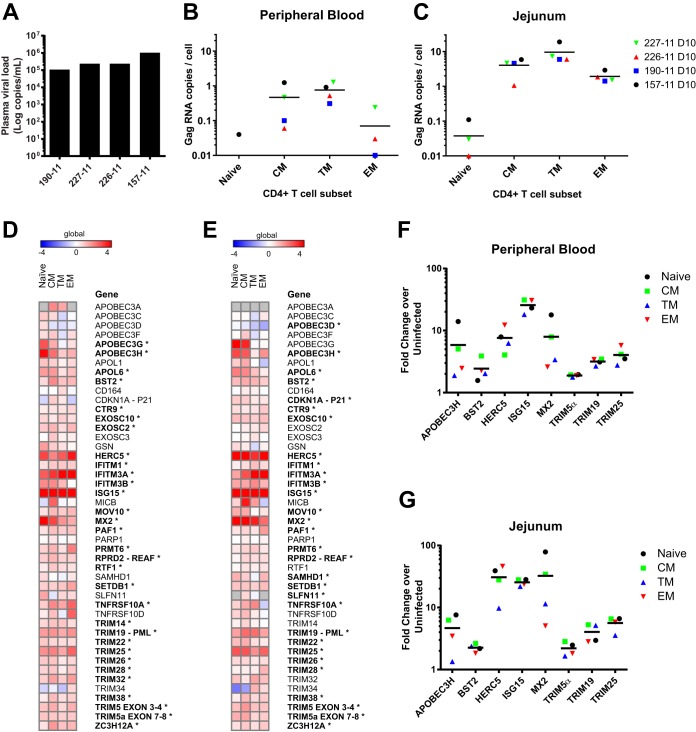
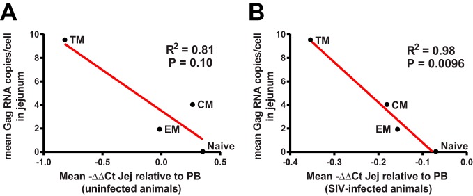
Although multiple restriction factors have been shown to inhibit HIV/SIV replication, little is known about their expression in vivo Expression of 45 confirmed and putative HIV/SIV restriction factors was analyzed in CD4+ T cells from peripheral blood and the jejunum in rhesus macaques, revealing distinct expression patterns in naive and memory subsets. In both peripheral blood and the jejunum, memory CD4+ T cells expressed higher levels of multiple restriction factors compared to naive cells. However, relative to their expression in peripheral blood CD4+ T cells, jejunal CCR5+ CD4+ T cells exhibited significantly lower expression of multiple restriction factors, including APOBEC3G, MX2, and TRIM25, which may contribute to the exquisite susceptibility of these cells to SIV infection. In vitro stimulation with anti-CD3/CD28 antibodies or type I interferon resulted in upregulation of distinct subsets of multiple restriction factors. After infection of rhesus macaques with SIVmac239, the expression of most confirmed and putative restriction factors substantially increased in all CD4+ T cell memory subsets at the peak of acute infection. Jejunal CCR5+ CD4+ T cells exhibited the highest levels of SIV RNA, corresponding to the lower restriction factor expression in this subset relative to peripheral blood prior to infection. These results illustrate the dynamic modulation of confirmed and putative restriction factor expression by memory differentiation, stimulation, tissue microenvironment and SIV infection and suggest that differential expression of restriction factors may play a key role in modulating the susceptibility of different populations of CD4+ T cells to lentiviral infection.IMPORTANCE Restriction factors are genes that have evolved to provide intrinsic defense against viruses. HIV and simian immunodeficiency virus (SIV) target CD4+ T cells. The baseline level of expression in vivo and degree to which expression of restriction factors is modulated by conditions such as CD4+ T cell differentiation, stimulation, tissue location, or SIV infection are currently poorly understood. We measured the expression of 45 confirmed and putative restriction factors in primary CD4+ T cells from rhesus macaques under various conditions, finding dynamic changes in each state. Most dramatically, in acute SIV infection, the expression of almost all target genes analyzed increased. These are the first measurements of many of these confirmed and putative restriction factors in primary cells or during the early events after SIV infection and suggest that the level of expression of restriction factors may contribute to the differential susceptibility of CD4+ T cells to SIV infection.

Keywords: CD4+ T lymphocytes; gut-associated lymphoid tissue; interferon-stimulated genes; restriction factors; simian immunodeficiency virus.


Figures

FIG 1
FIG 1
Confirmed and putative restriction factors exhibit similar patterns of expression in peripheral blood and jejunum CD4+ T cell memory subsets and are dynamically modulated by stimulation. (A and B) Heat map of −ΔΔCT expression values depicting memory subset expression relative to naive for peripheral blood (A) and the jejunum (B). POLR2A was used as endogenous control gene for normalization. The means for four animals are shown (*, P < 0.01 [repeated-measures ANOVA with an extension of the Benjamini-Hochberg correction method]). (C and D) PCA for peripheral blood (C) and the jejunum (D) based on analysis of 45 restriction factor genes. Each symbol in panels C and D represents a different animal. (E and F) PC1 loading factors from the PCA for peripheral blood (E) and the jejunum (F).
FIG 2
FIG 2
Expression of confirmed and putative restriction factors is dynamically modulated by CD3/CD28 and type I IFN stimulation. (A) Time course of the change in expression of selected restriction factor genes in CD4+ T cells from peripheral blood stimulated with anti-CD3/anti-CD28 beads versus mock-treated animals relative to the time of stimulation. (B) Time course of the change in expression of restriction factor genes after stimulation with 1,000 U of type I IFN/ml relative to the time of stimulation. Data represent the means of samples obtained from four animals. The results shown for TRIM5α reflect data obtained using an assay specific for the α isoform of TRIM5.
FIG 3
FIG 3
Differences in expression of confirmed and putative restriction factors in peripheral blood and jejunum CD4+ T cells. (A) Heat map depicting expression in the jejunum relative to peripheral blood and the most stable endogenous control gene, B2M. Unsupervised hierarchical clustering was performed using an uncentered Pearson correlation with complete linkage groups genes with similar expression patterns. The means of four animals are shown (*, P < 0.05 [two-way ANOVA testing tissue differences with an extension of the Benjamini-Hochberg correction method]). (B) Mean expression plus the standard deviations (−ΔΔCT) of all restriction factors in the jejunum relative to their respective peripheral blood subset (*, P < 0.05 [paired t test]).
FIG 4
FIG 4
Increased SIV infection in jejunum transitional memory CD4+ T cells and changes in expression of confirmed and putative restriction factors during acute infection. (A) Plasma viral load for each of the four SIV-infected animals at day 10 postinfection. (B and C) The Gag RNA copies per cell equivalent were quantified in sorted CD4+ T cell subsets from peripheral blood (B) and the jejunum (C). (D and E) Heat map of −ΔΔCT expression values depicting memory subset expression in postinfection cells relative to uninfected animals for peripheral blood (D) and the jejunum (E). B2M was used as an endogenous control gene to normalize both uninfected and infected data. Means of four animals per group are shown (*, P < 0.01 [two-way ANOVA with an extension of the Benjamini-Hochberg correction method testing differences due to infection]). (F and G) Mean fold change of selected restriction factors from CD4+ T cell subsets postinfection relative to the same subset in uninfected animals for peripheral blood (F) and the jejunum (G).
FIG 5
FIG 5
Correlation of expression of confirmed and putative restriction factors with the outcome of infection in subsets of CD4+ T cells. The relatively lower levels of expression of confirmed and putative restriction factors in CD4+ T cell subsets in the jejunum compared to peripheral blood (PB) in uninfected animals (ΔΔCT) (A) and in SIV-infected animals (B) correlated with the level of SIV infection in jejunal CD4+ T cell subsets. Data for each CD4+ subset represent the means of four uninfected animals and four infected animals. R2 and P values were calculated using a two-tailed Pearson correlation.

Similar articles

Cited by

References

    1. Sheehy AM, Gaddis NC, Malim MH. 2003. The antiretroviral enzyme APOBEC3G is degraded by the proteasome in response to HIV-1 Vif. Nat Med 9:1404–1407. doi:10.1038/nm945. - DOI - PubMed
    1. Stremlau M, Perron M, Lee M, Li Y, Song B, Javanbakht H, Diaz-Griffero F, Anderson DJ, Sundquist WI, Sodroski J. 2006. Specific recognition and accelerated uncoating of retroviral capsids by the TRIM5α restriction factor. Proc Natl Acad Sci U S A 103:5514–5519. doi:10.1073/pnas.0509996103. - DOI - PMC - PubMed
    1. Neil SJ, Zang T, Bieniasz PD. 2008. Tetherin inhibits retrovirus release and is antagonized by HIV-1 Vpu. Nature 451:425–430. doi:10.1038/nature06553. - DOI - PubMed
    1. Van Damme N, Goff D, Katsura C, Jorgenson RL, Mitchell R, Johnson MC, Stephens EB, Guatelli J. 2008. The interferon-induced protein BST-2 restricts HIV-1 release and is downregulated from the cell surface by the viral Vpu protein. Cell Host Microbe 3:245–252. doi:10.1016/j.chom.2008.03.001. - DOI - PMC - PubMed
    1. Laguette N, Sobhian B, Casartelli N, Ringeard M, Chable-Bessia C, Segeral E, Yatim A, Emiliani S, Schwartz O, Benkirane M. 2011. SAMHD1 is the dendritic- and myeloid-cell-specific HIV-1 restriction factor counteracted by Vpx. Nature 474:654–657. doi:10.1038/nature10117. - DOI - PMC - PubMed

Publication types

MeSH terms

LinkOut - more resources