. 2017 Mar 13;91(7):e00062-17.
doi: 10.1128/JVI.00062-17. Print 2017 Apr 1.

A Temperature-Sensitive Lesion in the N-Terminal Domain of the Rotavirus Polymerase Affects Its Intracellular Localization and Enzymatic Activity

Affiliations

A Temperature-Sensitive Lesion in the N-Terminal Domain of the Rotavirus Polymerase Affects Its Intracellular Localization and Enzymatic Activity

Allison O McKell et al. J Virol. .

Abstract

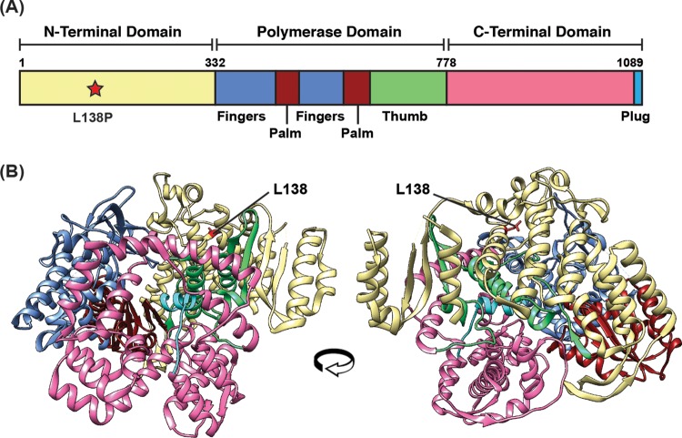
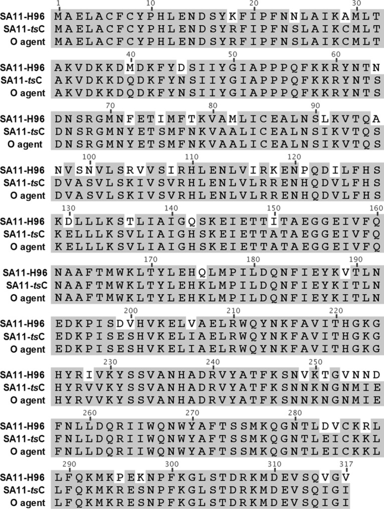
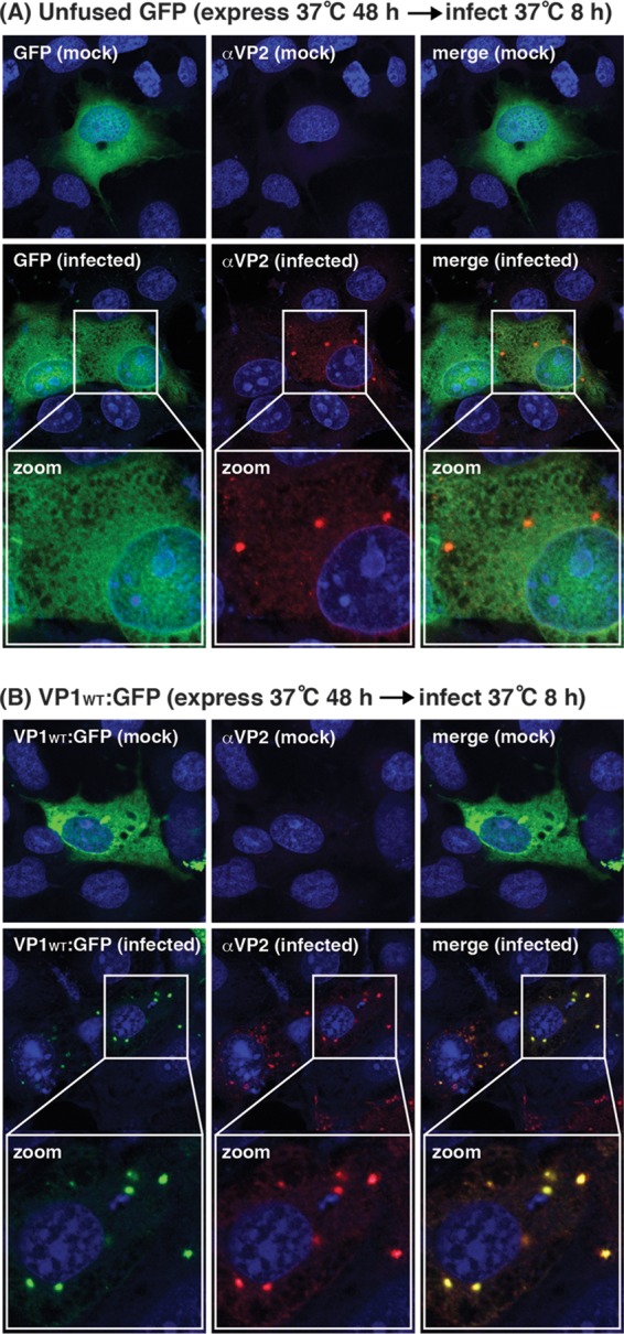
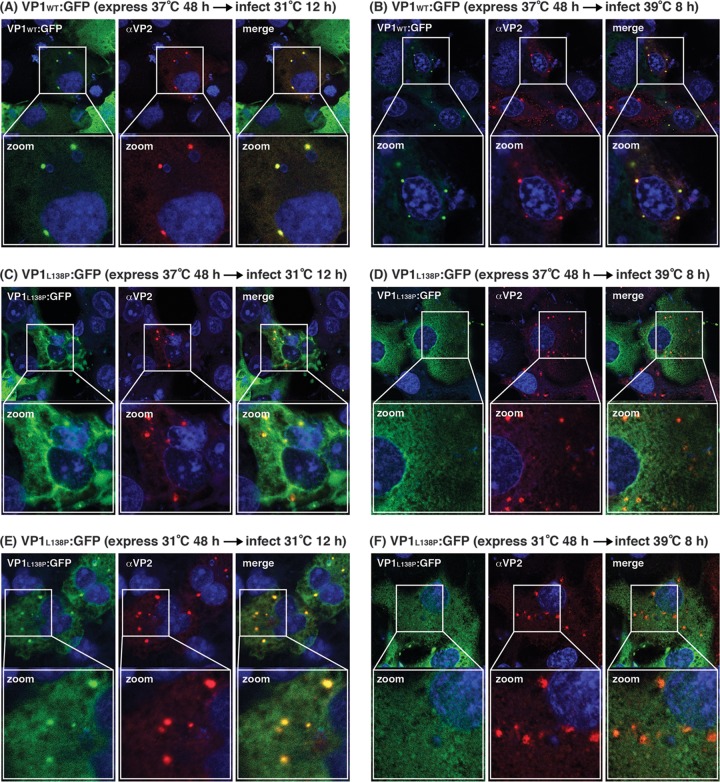
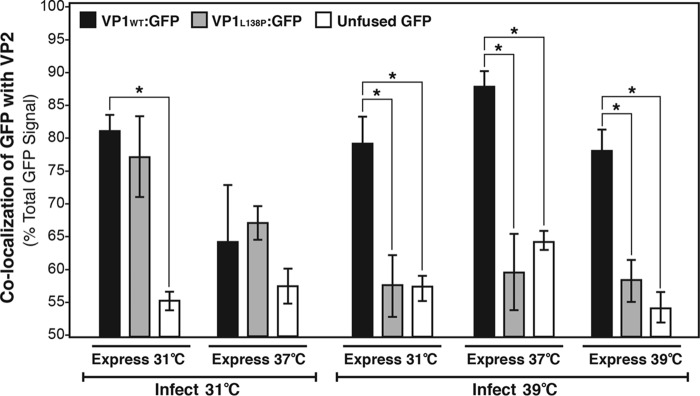
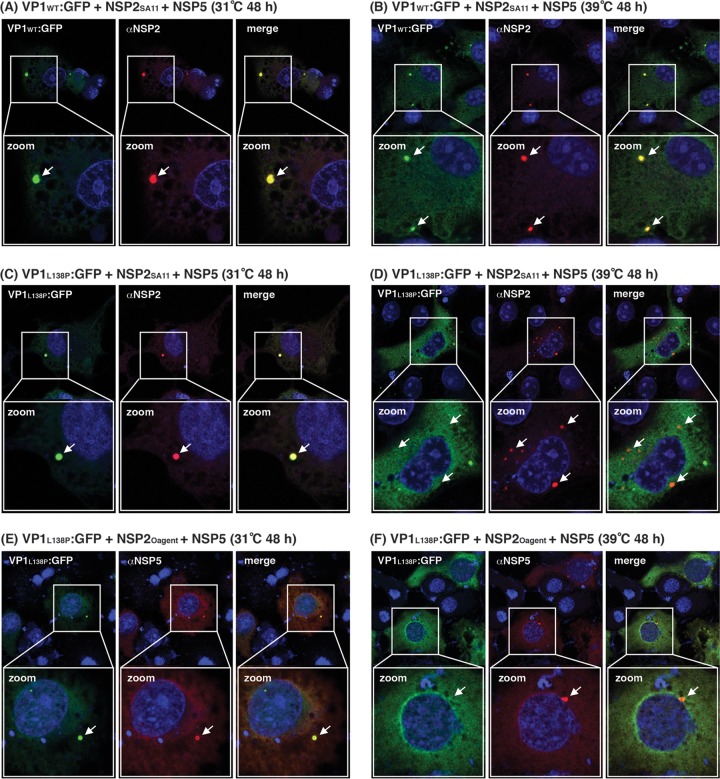
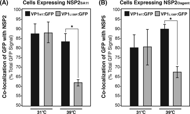
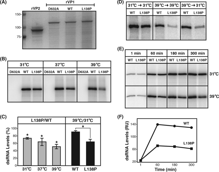
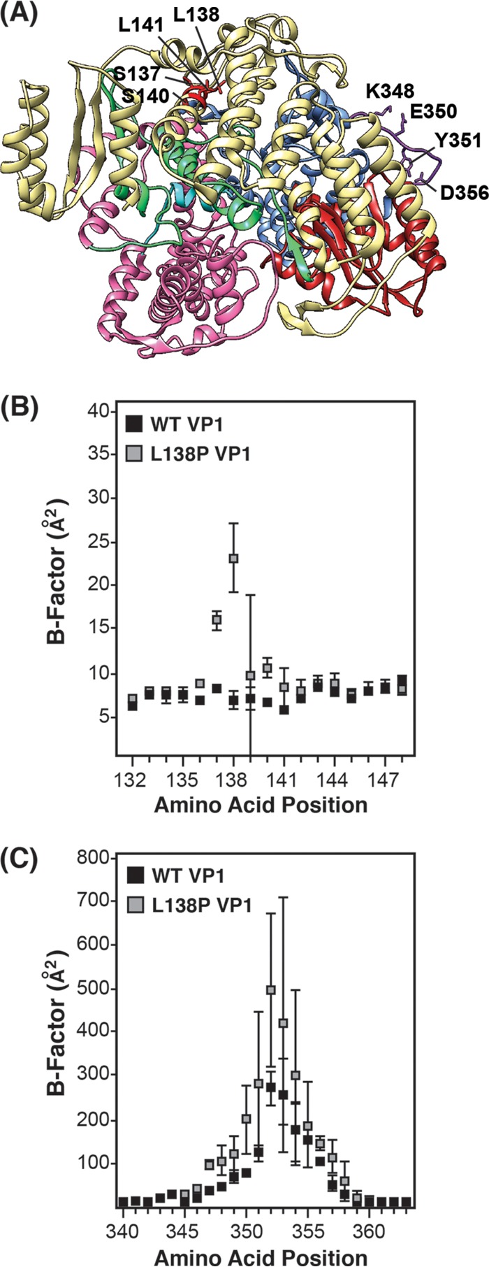
Temperature-sensitive (ts) mutants of simian rotavirus (RV) strain SA11 have been previously created to investigate the functions of viral proteins during replication. One mutant, SA11-tsC, has a mutation that maps to the gene encoding the VP1 polymerase and shows diminished growth and RNA synthesis at 39°C compared to that at 31°C. In the present study, we sequenced all 11 genes of SA11-tsC, confirming the presence of an L138P mutation in the VP1 N-terminal domain and identifying 52 additional mutations in four other viral proteins (VP4, VP7, NSP1, and NSP2). To investigate whether the L138P mutation induces a ts phenotype in VP1 outside the SA11-tsC genetic context, we employed ectopic expression systems. Specifically, we tested whether the L138P mutation affects the ability of VP1 to localize to viroplasms, which are the sites of RV RNA synthesis, by expressing the mutant form as a green fluorescent protein (GFP) fusion protein (VP1L138P-GFP) (i) in wild-type SA11-infected cells or (ii) in uninfected cells along with viroplasm-forming proteins NSP2 and NSP5. We found that VP1L138P-GFP localized to viroplasms and interacted with NSP2 and/or NSP5 at 31°C but not at 39°C. Next, we tested the enzymatic activity of a recombinant mutant polymerase (rVP1L138P) in vitro and found that it synthesized less RNA at 39°C than at 31°C, as well as less RNA than the control at all temperatures. Together, these results provide a mechanistic basis for the ts phenotype of SA11-tsC and raise important questions about the role of leucine 138 in supporting key protein interactions and the catalytic function of the VP1 polymerase.IMPORTANCE RVs cause diarrhea in the young of many animal species, including humans. Despite their medical and economic importance, gaps in knowledge exist about how these viruses replicate inside host cells. Previously, a mutant simian RV (SA11-tsC) that replicates worse at higher temperatures was identified. This virus has an amino acid mutation in VP1, which is the enzyme responsible for copying the viral RNA genome. The mutation is located in a poorly understood region of the polymerase called the N-terminal domain. In this study, we determined that the mutation reduces the ability of VP1 to properly localize within infected cells at high temperatures, as well as reduced the ability of the enzyme to copy viral RNA in a test tube. The results of this study explain the temperature sensitivity of SA11-tsC and shed new light on functional protein-protein interaction sites of VP1.

Keywords: RNA synthesis; VP1; polymerase; rotavirus; temperature-sensitive mutant; viroplasm.


Figures

FIG 1
FIG 1
VP1 domain organization and location of the L138P mutation. (A) Linear schematic of VP1 (not drawn to scale). The N-terminal (yellow) and C-terminal (pink) domains flank the central polymerase domain, which contains canonical finger (blue), palm (dark red), and thumb (green) subdomains. The extreme C terminus of the protein forms a plug subdomain (cyan). The amino acid residues making up each domain are listed at the top of the schematic, and the location of the L138P mutation in SA11-tsC is shown as a red star. (B) Atomic structure of VP1 (PDB accession no. 2R7R) in a ribbon representation. Domains and subdomains are colored as in panel A, and L138 is red and labeled. On the left side is the traditional front view of VP1, and on the right side is the protein rotated approximately 100° to the left so that L138 can be better visualized.
FIG 2
FIG 2
NSP2 amino acid sequence alignment. The primary NSP2 amino acid sequences of strains SA11-H96, SA11-tsC, and O agent are shown. Amino acid positions are listed at the top of the sequence alignment, and conserved residues are shaded gray.
FIG 3
FIG 3
Ectopically expressed WT VP1 localizes to viroplasms as a GFP fusion protein. Cos-7 cells on glass coverslips were transfected with plasmids expressing either unfused GFP (A) or the WT VP1 fusion protein VP1WT-GFP (B). Following their incubation at 37°C for 48 h, the cells were either mock infected or infected with SA11-4F (MOI of 10) at 37°C for 8 h. Immunofluorescence confocal microscopy was used to determine the localization of GFP (green) relative to viroplasms (anti-VP2; red) and nuclei (DAPI; blue). Colocalization of GFP and VP2 is yellow in the merged images.
FIG 4
FIG 4
Localization of VP1L138P-GFP in infected cells at various temperatures. Cos-7 cells on glass coverslips were transfected with plasmids expressing either VP1WT-GFP (A, B) or VP1L138P-GFP (C to F). Following their incubation for 48 h at the temperatures indicated, the cells were infected with SA11-4F (MOI of 10) at either 31°C for 12 h or 39°C for 8 h. Immunofluorescence confocal microscopy was used to determine the localization of GFP (green) relative to viroplasms (anti-VP2; red) and nuclei (DAPI; blue). Colocalization of GFP and VP2 is yellow in the merged images.
FIG 5
FIG 5
Quantitation of percent colocalization of GFP fusion proteins with VP2. Confocal micrographs of infected Cos-7 cells expressing VP1WT-GFP (black bars), VP1L138P-GFP (gray bars), or unfused GFP (white bars) were quantified for percent colocalization with viroplasms (i.e., anti-VP2 staining) at various temperatures with ImageJ software. Approximately four to six cells were quantified for each condition, and averages are shown. Error bars represent the standard deviation from the mean. Asterisks indicate P values of <0.01.
FIG 6
FIG 6
Localization of VP1L138P-GFP to viroplasm-like structures at various temperatures. Cos-7 cells on glass coverslips were transfected with plasmids expressing VP1WT-GFP, NSP2SA11, and NSP5 (A, B); VP1L138P-GFP, NSP2SA11, and NSP5 (C, D); and VP1L138P-GFP, NSP2Oagent, and NSP5 (E, F). The temperature and timing of protein expression are indicated. Immunofluorescence confocal microscopy was used to determine the localization of GFP (green) relative to viroplasm-like structures (anti-NSP2 or anti-NSP5; punctate red) and nuclei (DAPI; blue). Colocalization of GFP and NSP2/NSP5 is yellow in the merged images. The locations of punctate viroplasm-like structures formed of NSP2 and NSP5 are indicated by arrows in the zoomed images.
FIG 7
FIG 7
Quantitation of percent colocalization of GFP fusion proteins with viroplasm-like structures. Confocal micrographs of Cos-7 cells expressing either VP1WT-GFP (black bars) or VP1L138P-GFP (gray bars) along with NSP2 and NSP5 were quantified with ImageJ software for percent colocalization with NSP2 in cells expressing NSP2SA11 (A) or NSP5 in cells expressing NSP2Oagent (B). Approximately four to six cells were quantified for each condition, and averages are shown. Error bars represent the standard deviation from the mean. Asterisks indicate P values of <0.01.
FIG 8
FIG 8
In vitro dsRNA synthesis by recombinant VP1 proteins. (A) Purified recombinant polymerase protein (rVP1) or core shell protein (rVP2) was electrophoresed in an SDS-polyacrylamide gel and stained with GelCode Blue. Molecular mass standards (in kilodaltons) are shown to the left. WT and catalytically inactive (D632A mutant) proteins served as positive and negative controls, respectively, for the activity of the mutant (L138P) protein. (B) dsRNA synthesis by rVP1 proteins at various temperatures. Radiolabeled dsRNA products made by each rVP1 during 180-min reactions at 31°C, 37°C, or 39°C were electrophoresed in SDS-polyacrylamide gels and detected with a phosphorimager. (C) Quantification of dsRNA levels. The left portion of the graph shows the percentage of dsRNA made by rVP1L138P relative to that made by rVP1WT (L138P/WT). The right portion of the graph shows the dsRNA level made by each protein at 39°C as a percentage of that made at 31°C (39°C/31°C). Asterisks indicate P values of <0.01. (D) dsRNA synthesis by rVP1 proteins following a temperature shift. rVP1 proteins were incubated at either 31°C or 39°C for 60 min prior to being assayed for dsRNA synthesis at 31°C or 39°C for 180 min. dsRNA products were electrophoresed in SDS-polyacrylamide gels and detected with a phosphorimager. (E) Time course of dsRNA synthesis. Radiolabeled dsRNA products made by each rVP1 at the times and temperatures indicated were electrophoresed in SDS-polyacrylamide gels and detected with a phosphorimager. (F) Quantification of time course. Radiolabeled dsRNA from three independent experiments was quantified (relative units [RU]). The graph shows the results of a single representative experiment.
FIG 9
FIG 9
Molecular dynamics simulations of VP1 protein structures. (A) Structure of WT VP1 with modeled loop (residues 346 to 358). The view of WT VP1 is from the back, similar to that shown in Fig. 1B (right). Domains and subdomains are colored as in Fig. 1, except that the modeled loop is purple and the region proximal to L138 is bright red. Residues with significantly different B factors are shown as stick models and labeled. Average B factors are shown for the region proximal to position 138 (B) and the modeled loop region (C). In both panels B and C, residue position numbers are shown on the x axis, WT VP1 B factors are plotted as black boxes, and L138P mutant VP1 B factors are plotted as gray boxes. Error bars represent the standard deviation from the mean following three independent simulations.

Similar articles

Cited by

References

    1. Estes MK, Kapikian AZ. 2006. Rotaviruses and their replication, p 1917–1974. In Knipe DM, Howley PM (ed), Fields virology, 5th ed Lippincott Williams & Wilkins, Philadelphia, PA.
    1. Tate JE, Burton AH, Boschi-Pinto C, Parashar UD, World Health Organization-Coordinated Global Rotavirus Surveillance Network. 2016. Global, regional, and national estimates of rotavirus mortality in children <5 years of age, 2000–2013. Clin Infect Dis 62(Suppl 2):S96–S105. doi:10.1093/cid/civ1013. - DOI - PMC - PubMed
    1. Li Z, Baker ML, Jiang W, Estes MK, Prasad BV. 2009. Rotavirus architecture at subnanometer resolution. J Virol 83:1754–1766. doi:10.1128/JVI.01855-08. - DOI - PMC - PubMed
    1. Settembre EC, Chen JZ, Dormitzer PR, Grigorieff N, Harrison SC. 2011. Atomic model of an infectious rotavirus particle. EMBO J 30:408–416. doi:10.1038/emboj.2010.322. - DOI - PMC - PubMed
    1. Greenberg HB, Kalica AR, Wyatt RG, Jones RW, Kapikian AZ, Chanock RM. 1981. Rescue of noncultivatable human rotavirus by gene reassortment during mixed infection with ts mutants of a cultivatable bovine rotavirus. Proc Natl Acad Sci U S A 78:420–424. doi:10.1073/pnas.78.1.420. - DOI - PMC - PubMed

LinkOut - more resources