. 2017 Mar 1;198(5):2182-2190.
doi: 10.4049/jimmunol.1601480. Epub 2017 Jan 18.

Th22 Cells Form a Distinct Th Lineage from Th17 Cells In Vitro with Unique Transcriptional Properties and Tbet-Dependent Th1 Plasticity

Affiliations

Th22 Cells Form a Distinct Th Lineage from Th17 Cells In Vitro with Unique Transcriptional Properties and Tbet-Dependent Th1 Plasticity

Maximilian W Plank et al. J Immunol. .

Abstract

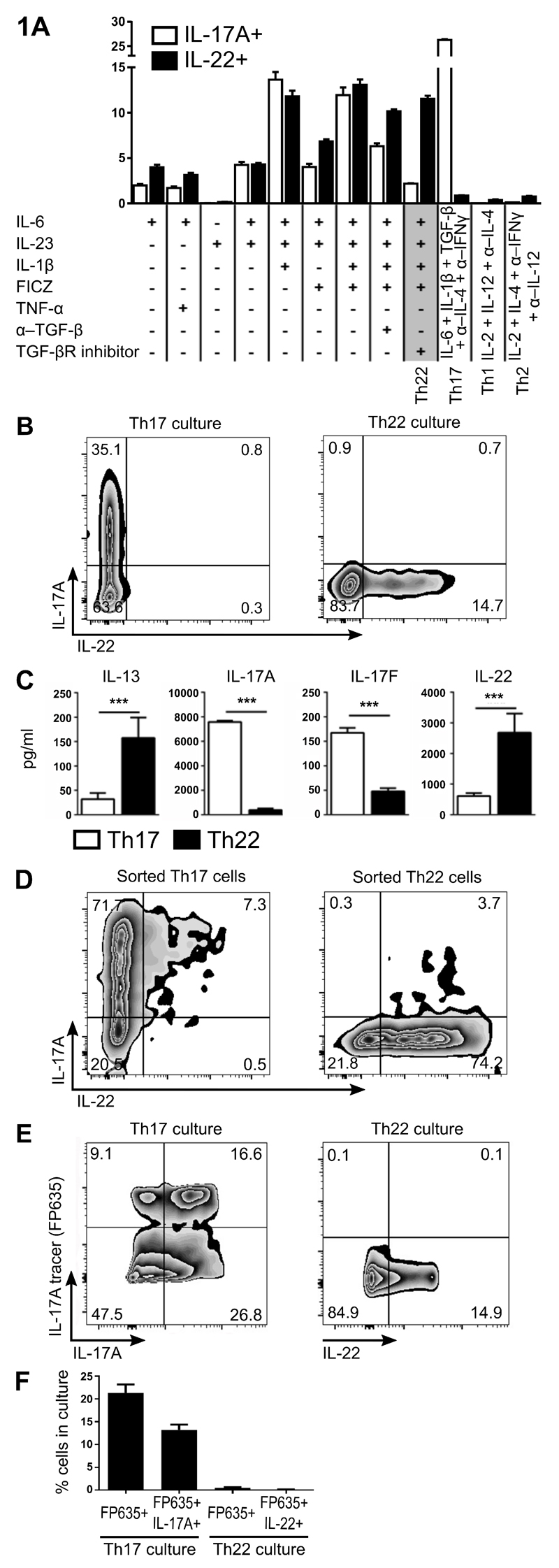
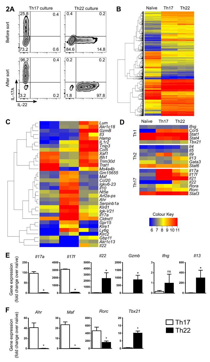
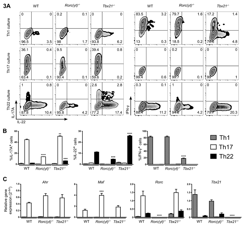
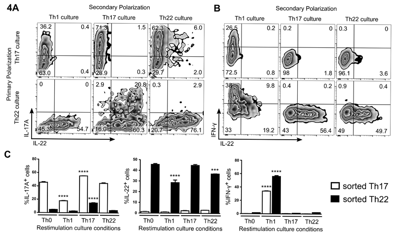
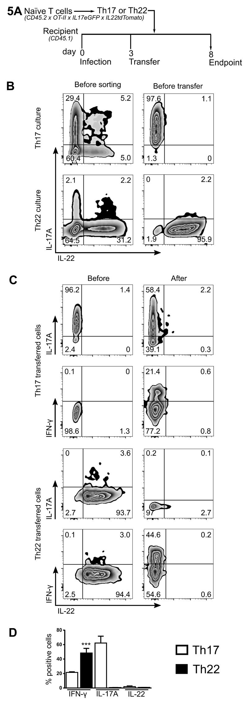
Th22 cells are a major source of IL-22 and have been found at sites of infection and in a range of inflammatory diseases. However, their molecular characteristics and functional roles remain largely unknown because of our inability to generate and isolate pure populations. We developed a novel Th22 differentiation assay and generated dual IL-22/IL-17A reporter mice to isolate and compare pure populations of cultured Th22 and Th17 cells. Il17a fate-mapping and transcriptional profiling provide evidence that these Th22 cells have never expressed IL-17A, suggesting that they are potentially a distinct cell lineage from Th17 cells under in vitro culture conditions. Interestingly, Th22 cells also expressed granzymes, IL-13, and increased levels of Tbet. Using transcription factor-deficient cells, we demonstrate that RORγt and Tbet act as positive and negative regulators of Th22 differentiation, respectively. Furthermore, under Th1 culture conditions in vitro, as well as in an IFN-γ-rich inflammatory environment in vivo, Th22 cells displayed marked plasticity toward IFN-γ production. Th22 cells also displayed plasticity under Th2 conditions in vitro by upregulating IL-13 expression. Our work has identified conditions to generate and characterize Th22 cells in vitro. Further, it provides evidence that Th22 cells develop independently of the Th17 lineage, while demonstrating plasticity toward both Th1- and Th2-type cells.


Figures

Figure 1
Figure 1. Generation of Th22 cells that appears to be a distinct lineage to Th17 cells
Determination of optimal culture conditions for Th22 cells (IL-1β, IL-6, IL-23, FICZ and TGF-βR inhibitor/Galunisertib, with anti-IL-4/IFNγ). Percentage of IL-17A+ and IL-22+ cells after culturing naïve Th cells under various polarizing conditions are graphed (A). Representative plots of naïve Th cells stimulated for 3 d under optimal Th17 conditions or Th22 conditions (B). Cytokine protein levels in culture SN from naïve Th cells stimulated for 3 d under optimal Th17 conditions or Th22 conditions (C). Naïve Th cells from IL-17eGFP x IL-22tdTomato reporter mice were differentiated with Th17 or Th22 conditions then FACS sorted to purify Th17 or Th22, respectively. Representative plots of FACS-sorted Th17 and Th22 cells re-stimulated for 3 d under Th0 conditions (D). Representative plots (E), and percentage quantitation (F) of IL-17A fate mapping reporter (IL-17A tracer: Il17aFP635) expression versus IL-17A or IL-22 protein expression in naïve Th cells differentiated for 3 d under optimal Th17 or Th22 conditions. Cell populations in FACS plots are pre-gated on CD4+CD44+ and viable cells. Error bars represent SEM (n=6 per group from three independent experiments). ***p<0.001.
Figure 2
Figure 2. Transcription profiling in sorted pure Th17 and Th22 cells.
Cultures of enriched Th17 and Th22 cells were generated from IL-17eGFP x IL-22tdTomato reporter mice using optimal polarizing conditions for the first 3 d, and cells sorted on day 4 for Th17 cells (CD4+CD44+IL-17eGFP+) or Th22 cells (CD4+CD44+IL-17eGFP-IL-22tdTomato+). RNA was extracted from sorted cells and mRNA analysis performed. Th17 and Th22 enriched cultures before FACS, and resulting populations after cell sorting (viable cells shown) (A). Overview heatmap showing differentially expressed mRNAs in sorted naïve Th, Th17 and Th22 cells (B). Heatmap with 33 differentially regulated genes between Th22 and Th17 cells (>6-fold) shown for naïve Th, Th17 and Th22 cells (C). Th cell signature genes (Th1, Th2 and Th17) shown for naïve Th, Th17 and Th22 cells (D). RT-PCR confirmation of cytokine expression (E) and transcription factors (F) in sorted Th17 and Th22 cells (expression is normalized to naïve T cells). Error bars represent SEM (n=6 per group from three independent experiments). *p < 0.05.
Figure 3
Figure 3. Transcription factor control of Th17 and Th22 cell differentiation.
Representative plots of WT, Rorc(γt)-/- and Tbx21-/- Th cells differentiated in each Th condition (Th1, Th17 and Th22) (A). Cell populations in FACS plots are pre-gated on CD4+CD44+ and viable cells. Quantitation of IL-17+, IL-22+ and IFN-γ+ cells under each Th condition (Th1, Th17 and Th22) for WT, Rorc(γt)-/- and Tbx21-/- cells (B) and differential transcription factor expression assessed by RT-PCR, normalized to the housekeeping gene Hprt (C). Error bars represent SEM (n=6 per group from three independent experiments). **p < 0.01, ***p < 0.001, ****p < 0.0001.
Figure 4
Figure 4. In vitro plasticity of sorted Th22 cells re-stimulated under Th1, Th17 and Th22 conditions.
Cultures of enriched Th17 and Th22 cells were generated from IL-17eGFP x IL-22tdTomato reporter mice, using optimal polarizing conditions for the first 3 d, and cells sorted on day 4 for Th17 cells (CD4+CD44+IL-17eGFP+) or Th22 cells (CD4+CD44+IL-17eGFP-IL-22tdTomato+). Purified Th17 and Th22 cells were re-stimulated under Th1, Th17 and Th22 conditions for a further 3 d and the expression of IL-17A, IL-22 and IFN-γ determined. Representative plots of IL-17eGFP and IL-22tdTomato expression in re-stimulated, unfixed Th17 and Th22 cells are shown (A). Representative plots of IL-17eGFP, IL-22tdTomato, and IFN-γ expression in re-stimulated, fixed Th17 and Th22 cells are shown (B). Cell populations in FACS plots are pre-gated on CD4+CD44+ and viable cells. Quantification of IL-17eGFP+, IL-22tdTomato+ and IFN-γ+ cells in sorted Th17 and Th22 populations re-stimulated under Th0, Th1, Th17 and Th22 conditions (C). Note that paraformaldehyde fixation interferes with fluorescence from the tdTomato protein and likely explains the observed decrease in tdTomato signal following intracellular staining (Figure 4B/C). Error bars represent SEM (n=6 per group from three independent experiments). ***p < 0.001, ****p < 0.0001.
Figure 5
Figure 5. In vivo plasticity of Th22 cells in a high IFN/Th1-inflammatory environment.
Naïve Th cells from IL-17eGFP x IL-22tdTomato x OT-II TCR-transgenic (CD45.2) mice were used to differentiate antigen-specific (ovalbumin323–339) Th17 and Th22 cells. OVA-specific Th17 and Th22 cells were purified by FACS-sorting before being transferred into recipient mice (CD45.1) on day 3. Recipient animals were infected with Influenza A (1.4 x 10⁴ pfu of ovalbumin323–339 peptide-expressing influenza A virus; A/HK/x31; H3N2) 3 d before transfer. Endpoint analysis was performed 5 d after cell transfer. Model schematic (A). Representative plots of IL-17eGFP and IL-22tdTomato expression in CD45.2+ Th17 and Th22 enriched cultures before sorting, before adoptive transfer on day 3 (i.e. sorted Th17 and Th22 cells) (B). Representative plots of IL-17eGFP, IL-22tdTomato, and IFN-γ expression in CD45.2+ Th17 and Th22 cells before transfer, and in those cells isolated from the lungs 5 d after transfer into Influenza A-infected recipient mice (C). All cell populations in FACS plots are pre-gated on viable cells and after adoptive transfer additionally with CD4+CD45.2+. Quantitation of IL-17eGFP, IL-22tdTomato, and IFN-γ expression in CD45.2+ Th17 and Th22 cells 5 d after transfer into Influenza A-infected recipient mice (D). Error bars represent SEM (n=6 per group from three independent experiments). ***p<0.001.

References

    1. Kara EE, Comerford I, Fenix KA, Bastow CR, Gregor CE, McKenzie DR, McColl SR. Tailored immune responses: novel effector helper T cell subsets in protective immunity. PLoS pathogens. 2014;10(2):e1003905. - PMC - PubMed
    1. Li P, Spolski R, Liao W, Leonard WJ. Complex interactions of transcription factors in mediating cytokine biology in T cells. Immunological reviews. 2014;261(1):141–156. - PMC - PubMed
    1. Chung Y, Yang X, Chang SH, Ma L, Tian Q, Dong C. Expression and regulation of IL-22 in the IL-17-producing CD4+ T lymphocytes. Cell Res. 2006;16(11):902–907. - PubMed
    1. Liang SC, Tan XY, Luxenberg DP, Karim R, Dunussi-Joannopoulos K, Collins M, Fouser LA. Interleukin (IL)-22 and IL-17 are coexpressed by Th17 cells and cooperatively enhance expression of antimicrobial peptides. J Exp Med. 2006;203(10):2271–2279. - PMC - PubMed
    1. Zenewicz LA, Yancopoulos GD, Valenzuela DM, Murphy AJ, Stevens S, Flavell RA. Innate and adaptive interleukin-22 protects mice from inflammatory bowel disease. Immunity. 2008;29(6):947–957. - PMC - PubMed

MeSH terms