. 2017 Feb;216(2):463-476.
doi: 10.1083/jcb.201605110. Epub 2017 Jan 18.

Heat stress induces ferroptosis-like cell death in plants

Affiliations

Heat stress induces ferroptosis-like cell death in plants

Ayelén Mariana Distéfano et al. J Cell Biol. 2017 Feb.

Abstract

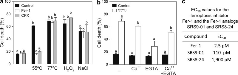
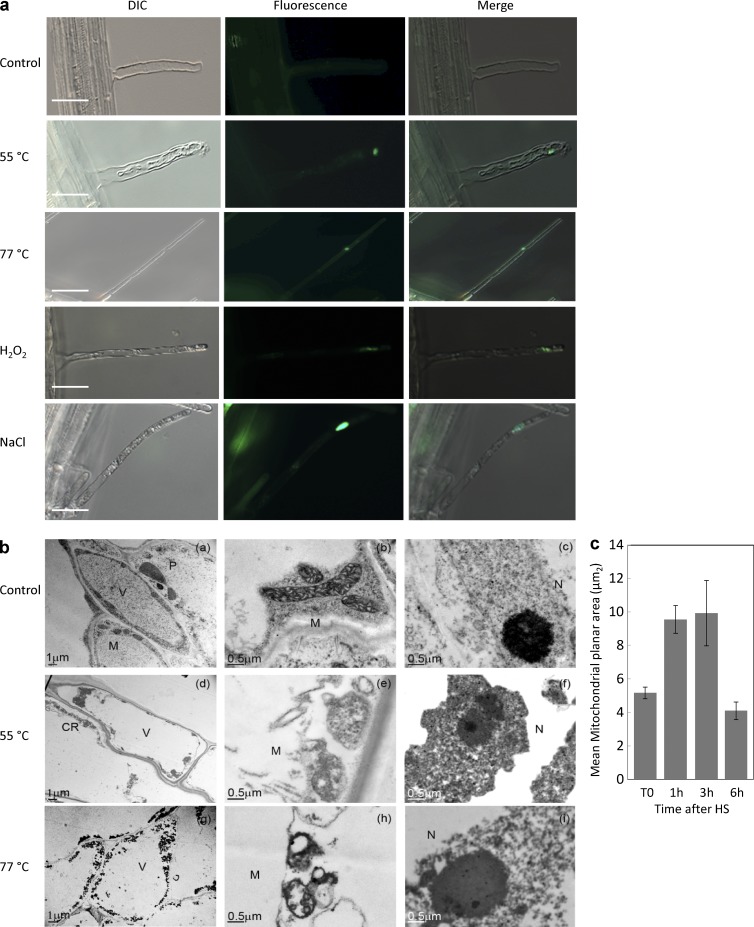
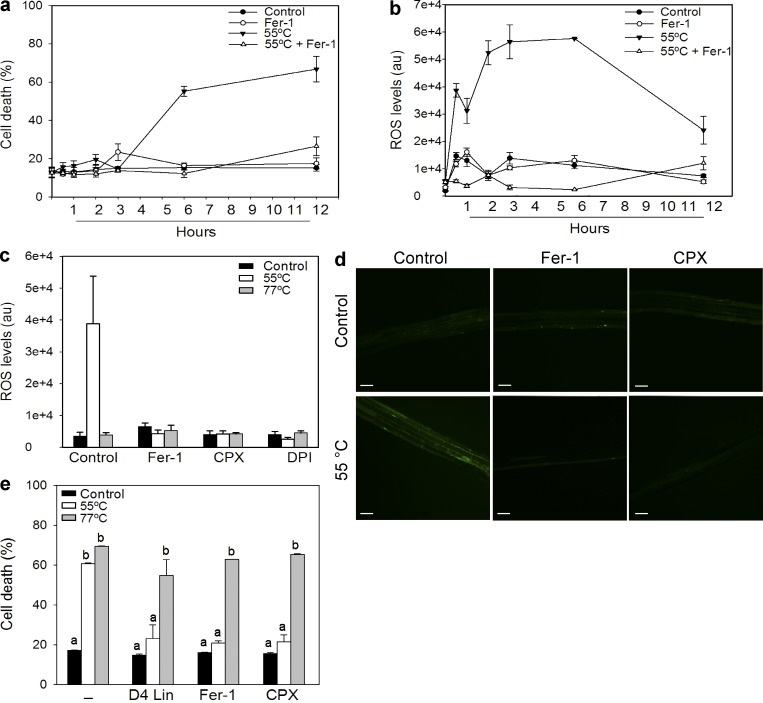
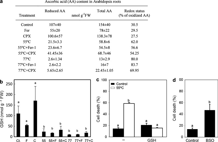
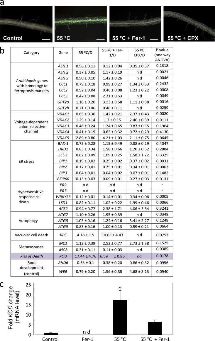
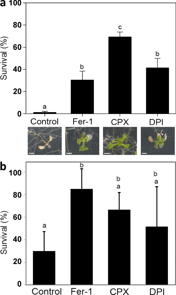
In plants, regulated cell death (RCD) plays critical roles during development and is essential for plant-specific responses to abiotic and biotic stresses. Ferroptosis is an iron-dependent, oxidative, nonapoptotic form of cell death recently described in animal cells. In animal cells, this process can be triggered by depletion of glutathione (GSH) and accumulation of lipid reactive oxygen species (ROS). We investigated whether a similar process could be relevant to cell death in plants. Remarkably, heat shock (HS)-induced RCD, but not reproductive or vascular development, was found to involve a ferroptosis-like cell death process. In root cells, HS triggered an iron-dependent cell death pathway that was characterized by depletion of GSH and ascorbic acid and accumulation of cytosolic and lipid ROS. These results suggest a physiological role for this lethal pathway in response to heat stress in Arabidopsis thaliana The similarity of ferroptosis in animal cells and ferroptosis-like death in plants suggests that oxidative, iron-dependent cell death programs may be evolutionarily ancient.


Figures

Figure 1.
Figure 1.
Ferroptosis inhibitors prevent PCD induced by 55°C HS in Arabidopsis root hairs. (a) 6-d-old seedlings were preincubated with 1 µM Fer-1 (white bars), 10 µM CPX (gray bars), or DMSO (black bars). Cell death was induced by treating roots at 55°C or 77°C for 10 min, with H2O2 for 6 h, or with NaCl for 16 h. (b) 6-d-old seedlings were preincubated with CaCl2 for 16 h, with 1 mM EGTA for 2 h, or with EGTA for 2 h and then with CaCl2 for 16 h before inducing cell death by treating roots at 55°C for 10 min. (a and b) Root hairs were stained with Sytox green, and Sytox-positive cells (interpreted as dead cells) and Sytox-negative cells were quantified. Results are expressed as a percentage of dead cells. Data are the mean + SEM of three independent experiments. Bars with different letters denote statistical difference (one-way analysis of variance, P < 0.05). Also see Fig. S1. (c) 6-d-old seedlings were preincubated with Fer-1 analogues SR9-01 and SRS8-24 before treatment at 55°C. Root hairs were stained with Sytox green, and the number of Sytox-positive cells (interpreted as dead cells) and Sytox-negative cells was quantified to obtain the EC50 of those compounds.
Figure 2.
Figure 2.
Morphology of root cells after inducing cell death. (a) 6-d-old seedlings were treated with DMSO (control), 55°C, or 77°C for 10 min or with H2O2 for 6 h or NaCl for 16 h. Root hairs were stained with Sytox green 6 h after HS or right after H2O2 or NaCl treatments. The appearance of cells was examined using DIC microscopy or fluorescent microscopy. Bars, 50 µm. (b) TEM micrographs of Arabidopsis root cortical cells 6 h after HS: (a–c), root cells from a nonstressed (control) root; (d–f) root cells from a 55°C-treated root, showing cytoplasmic retraction and shrunken, abnormal mitochondria; and (g–i) root cells from a 77°C-treated root, showing mitochondria with a light matrix and very few cristae. V, vacuole; N, nuclei; M, mitochondria; P, plastids; CR, cytoplasmic retraction. (c) Size of mitochondria in roots of plants submitted to HS. Roots were stained with MitoTracker green FM, and mitochondrial images were obtained using confocal microscopy at the times indicated. Quantification of mitochondrial area was performed using the Mito-Morphology macro for ImageJ software (T0, n = 93; 2 h, n = 58; 3 h, n = 33; 6 h, n = 62).
Figure 3.
Figure 3.
Ferroptosis inhibitors prevent cell death and ROS accumulation induced by HS in Arabidopsis roots. 6-d-old seedlings were preincubated with 1 µM Fer-1, 10 µM CPX, or DMSO (Control) as indicated. Cell death was induced by treating roots at 55°C for 10 min. (a) Kinetics of cell death induced by a 55°C HS. (b) Kinetics of ROS levels induced after a 55°C HS treatment. ROS accumulation in roots was detected by using the H2DCFDA probe. (c) ROS production is mediated by NOX activity and prevented by cotreatment with ferroptosis inhibitors or cotreatment with the NOX inhibitor DPI. Data are the mean ± SEM of three independent experiments. (d) Representative confocal images showing ROS detected by the H2DCFDA probe in A. thaliana roots. Bars, 50 µm. (e) Suppression of cell death after a 55°C HS by D4-linoleate (D4 Lin). 6-d-old seedlings were preincubated with 1 µM Fer-1, 10 µM CPX, 8 µM D4-linoleate, or DMSO (control) as indicated. Cell death was induced by treating roots at 55°C or 77°C for 10 min. (a and e) Root hairs were stained with Sytox green, and Sytox-positive (interpreted as dead cells) and Sytox-negative cells were quantified. Results are expressed as a percentage of dead cells. Bars with different letters denote statistical difference (one-way analysis of variance, P < 0.05).
Figure 4.
Figure 4.
Plant ferroptosis involves GSH and AA oxidation and depletion. (a) AA levels in A. thaliana roots before or after treatment at 55°C or 77°C with or without the ferroptosis inhibitors Fer-1 and CPX. FW, fresh weight. (b) GSH levels in A. thaliana roots before (Ct., control) or after treatment at 55°C (55) or at 77°C with or without the ferroptosis inhibitors Fer-1 (F) and CPX (C). Data are the mean ± SEM of three independent experiments. Different letters denote statistical difference (one-way analysis of variance, P < 0.05). (c) 6-d-old seedlings were preincubated with 100 µM GSH or DMSO overnight (16 h). Cell death was induced by treating roots at 55°C. (d) Arabidopsis seedlings were grown in control plates or 10 µM l-buthionine-(S,R)-sulfoximine (BSO)–supplemented plates. 6-d-old seedlings were analyzed. (c and d) Root hairs were stained with Sytox green, and Sytox-positive (interpreted as dead cells) and Sytox-negative cells and the number of dead and living root hairs was quantified. Results are expressed as a percentage of dead cells. Data are mean ± SEM of three independent experiments. Different letters denote statistical difference (one-way analysis of variance, P < 0.05).
Figure 5.
Figure 5.
Expression of candidate Arabidopsis ferroptosis marker genes. (a) Arabidopsis roots from 6-d-old seedlings were preincubated with 1 µM Fer-1, 10 µM CPX, or DMSO (control). Cell death was induced by treating roots at 55°C for 10 min, and roots were stained with Sytox green 6 h after treatment. The images show that cells in the main root undergo cell death that is prevented by ferroptosis inhibitors. Bars, 50 µm. (b) Changes in expression of candidate genes that might be associated with plant ferroptosis in A. thaliana roots 2 h after HS at 55°C for 10 min. mRNA levels are expressed as a fold-change ratio between the different conditions and were analyzed by one-way analysis of variance (ANOVA). D, DMSO; nd, not determined. (c) mRNA expression levels of KOD determined by real-time quantitative PCR in A. thaliana roots in response to 55°C for 2 h. Data are from three independent biological replicates and presented as mean ± SD. Data were analyzed by one-way analysis of variance. *, P < 0.05. Significance is indicated relative to the treatment at 55°C plus the ferroptosis inhibitor Fer-1.
Figure 6.
Figure 6.
Ferroptosis inhibitors and DPI increase the rate of thermotolerance in Arabidopsis seedlings. (a) A. thaliana seedlings were grown on agar plates at 22°C for 6 d. Plates were pretreated as indicated for 16 h, exposed to HS (43°C for 1 h), and then returned to 22°C. The survival rate was determined 5 d after HS. Bottom panels show a representative picture of each case. Bars, 50 mm. (b) A. thaliana seedlings were grown on agar plates at 22°C for 6 d. Plates were pretreated as indicated in the dark for 16 h, exposed to HS (43°C for 1 h), and then returned to 22°C, avoiding exposure to light. The survival rate was determined 7 d after HS. (a and b) Control plates were pretreated with 1:1,000 DMSO. Each value is the mean ± SEM of at least 15 independent experiments. For each experiment, 20 seedlings per plate were tested. Different letters denote statistical difference (one-way analysis of variance, P < 0.05).

Comment in

  • Back to the roots of regulated necrosis.
    Todorov V, Linkermann A. Todorov V, et al. J Cell Biol. 2017 Feb;216(2):303-304. doi: 10.1083/jcb.201612078. Epub 2017 Jan 20. J Cell Biol. 2017. PMID: 28108525 Free PMC article.
  • Ferroptosis-like cell death in plants.
    Mushegian AA. Mushegian AA. Sci Signal. 2017 Feb 28;10(468):eaan0450. doi: 10.1126/scisignal.aan0450. Sci Signal. 2017. PMID: 28246195
  • Ferroptosis: Yet Another Way to Die.
    Kazan K, Kalaipandian S. Kazan K, et al. Trends Plant Sci. 2019 Jun;24(6):479-481. doi: 10.1016/j.tplants.2019.03.005. Epub 2019 Mar 23. Trends Plant Sci. 2019. PMID: 30910286

References

    1. Bartoli C.G., Yu J., Gómez F., Fernández L., McIntosh L., and Foyer C.H.. 2006. Inter-relationships between light and respiration in the control of ascorbic acid synthesis and accumulation in Arabidopsis thaliana leaves. J. Exp. Bot. 57:1621–1631. 10.1093/jxb/erl005 - DOI - PubMed
    1. Bergsbaken T., Fink S.L., and Cookson B.T.. 2009. Pyroptosis: Host cell death and inflammation. Nat. Rev. Microbiol. 7:99–109. 10.1038/nrmicro2070 - DOI - PMC - PubMed
    1. Bita C.E., and Gerats T.. 2013. Plant tolerance to high temperature in a changing environment: Scientific fundamentals and production of heat stress-tolerant crops. Front. Plant Sci. 4:273 10.3389/fpls.2013.00273 - DOI - PMC - PubMed
    1. Blanvillain R., Young B., Cai Y.M., Hecht V., Varoquaux F., Delorme V., Lancelin J.-M., Delseny M., and Gallois P.. 2011. The Arabidopsis peptide kiss of death is an inducer of programmed cell death. EMBO J. 30:1173–1183. 10.1038/emboj.2011.14 - DOI - PMC - PubMed
    1. Bollhöner B., Prestele J., and Tuominen H.. 2012. Xylem cell death: Emerging understanding of regulation and function. J. Exp. Bot. 63:1081–1094. 10.1093/jxb/err438 - DOI - PubMed

Publication types

MeSH terms