. 2017 Apr 20;45(7):4231-4243.
doi: 10.1093/nar/gkw1362.

Insights into the RecQ helicase mechanism revealed by the structure of the helicase domain of human RECQL5

Affiliations

Insights into the RecQ helicase mechanism revealed by the structure of the helicase domain of human RECQL5

Joseph A Newman et al. Nucleic Acids Res. .

Abstract

RecQ helicases are important maintainers of genome integrity with distinct roles in almost every cellular process requiring access to DNA. RECQL5 is one of five human RecQ proteins and is particularly versatile in this regard, forming protein complexes with a diverse set of cellular partners in order to coordinate its helicase activity to various processes including replication, recombination and DNA repair. In this study, we have determined crystal structures of the core helicase domain of RECQL5 both with and without the nucleotide ADP in two distinctly different ('Open' and 'Closed') conformations. Small angle X-ray scattering studies show that the 'Open' form of the protein predominates in solution and we discuss implications of this with regards to the RECQL5 mechanism and conformational changes. We have measured the ATPase, helicase and DNA binding properties of various RECQL5 constructs and variants and discuss the role of these regions and residues in the various RECQL5 activities. Finally, we have performed a systematic comparison of the RECQL5 structures with other RecQ family structures and based on these comparisons we have constructed a model for the mechano-chemical cycle of the common catalytic core of these helicases.


Figures

Figure 1.
Figure 1.
Structure of APO and ADP bound RECQL5. (A) Cartoon diagram of the APO (left) and ADP bound (right) RECQL5 forms with secondary structural elements colored by domain and the Zn2+ ion shown as a grey sphere. The top panel shows a schematic view of the domain arrangement in full length RECQL5. (B) Comparison of the Zn2+ binding subdomain across the RecQ family, the helical hairpin in RECQL5 is significantly longer than in other family members. (C) Close up view of the nucleotide binding site in the ADP form crystals, the ADP is shown is stick representation with polar contacts shown as black dashed lines and the 2Fo – 1Fc electron density map (contoured at 1σ) shown in grey covering the ADP/Mg2+.
Figure 2.
Figure 2.
Comparisons of various RECQL5 crystal forms. (A) Close up view of the active site showing a possible structural transition of motifs I, II, IV and the AR loop following nucleotide binding. (B) Comparison of the conformation of the six various chains of the monoclinic and triclinic ADP form crystals. (C) Close up view of the AR loop which was found to occupy two distinct states in the ADP form crystals, with H167 forming water mediated interactions with either Y419 (part of the Zn2+ domain, gray carbons) or T194 (part of motif III, blue carbons).
Figure 3.
Figure 3.
SAXS analysis of RECQL5 in solution. (A) Comparison of the theoretical (APO form in green and ADP form in red) and experimental scattering curves for the RECQL5A 11–453 construct, the experimental data shown was collected in the absence of nucleotide at 2 mg/ml. (B) SAXS scattering curves of the various length RECQL5 constructs follow a broadly similar profile. P(r) distance distribution functions (shown in the inset), and ab initio shape reconstructions shown in panel (C), are consistent with an increasingly elongated particle with similar cross sectional area.
Figure 4.
Figure 4.
Role of the RECQL5 ‘wedge’ helix in DNA binding. (A) Model of possible RECQL5 DNA complex based on a structural superposition with the BLM DNA complex, residues in the C-terminal ‘wedge’ helix that may contribute to DNA binding are labelled and shown in the stick format. (B) Characterization of RECQL5 11–609 DNA binding affinity and specificity by fluorescence polarization, showing a clear preference for binding splayed duplex, looped or single stranded DNA. (C) The RECQL5 11–438 construct lacking the ‘wedge’ helix had significantly reduced affinity and specificity. For all graphs, error bars are plotted as ±S.E. from three replicates. In the case of the 11–438 data, curves for all substrates were fitted with a single shared Bmax to ensure a more robust fit.
Figure 5.
Figure 5.
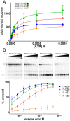
Characterization of the ATPase and Helicase activities of the various RECQL5 constructs. (A) DNA stimulated ATPase activity of RECQL5 constructs plotted as a function of ATP concentration. (B) Helicase activity of RECQL5 constructs, the upper panel shows representative gels and the lower panel shows quantification based on three independent experiments with error bars are plotted as ±S.E.
Figure 6.
Figure 6.
Characterization of the DNA binding, ATPase and helicase activities of RECQL5 variants. (A) DNA binding activity of RECQL5 variants binding to a splayed duplex DNA probe, only variants with significant difference from WT are shown for clarity. (B) DNA stimulated ATPase activity of RECQL5 variants plotted as a function of ATP concentration. (C) Helicase activity of RECQL5 variants, the upper panel shows representative gels and the lower panel shows quantification based on three independent experiments. The symbol scheme is shown on the bottom left hand side of panel and was used for all three plots and graphs are plotted as mean ±S.E. from three replicate experiments.
Figure 7.
Figure 7.
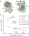
Quantitative comparison of the various RecQ family structures. (A) Multiple structural superposition of the current RecQ family structures aligned on the basis of the D1 domain (shown on the left) or the D2 domain (shown on the right) reveal structurally invariant positions from which to measure the relative domain positions. (B) 2D Plot of the D1–D2 conformation of the various RecQ family members as a function of the length of vectors between the invariant points. Individual chains of the same entry are shown separately where significant differences exist in their orientations.
Figure 8.
Figure 8.
RECQL5 helicase mechanism. Helicase activity is believed to be derived by switching between distinct conformational states driven successively by ATP binding, hydrolysis, Pi release and ADP release. The AR loop in the D1 domain is capable of both modulating the affinity of the D1 domain to DNA and stimulating ATPase activity. The AR loop conformation is sensitive to polar contacts to motif IV on the D2 domain and the nucleotide/Mg2+ binding status. Panel (A) shows an overview of the entire cycle with each of the four conformational states being linked to a representative PDB entry. Panel (B) shows a detailed view of the changes and transitions of the conserved catalytic helicase motifs in response to ATP binding, hydrolysis and release.

References

    1. Bachrati C.Z., Hickson I.D.. RecQ helicases: suppressors of tumorigenesis and premature aging. Biochem. J. 2003; 374:577–606. - PMC - PubMed
    1. German J., Archibal R., Bloom D.. Chromosomal breakage in a rare and probably genetically determined syndrome of man. Science. 1965; 148:506. - PubMed
    1. Yu C.E., Oshima J., Fu Y.H., Wijsman E.M., Hisama F., Alisch R., Matthews S., Nakura J., Miki T., Ouais S. et al. . Positional cloning of the Werner's syndrome gene. Science. 1996; 272:258–262. - PubMed
    1. Kitao S., Shimamoto A., Goto M., Miller R.W., Smithson W.A., Lindor N.M., Furuichi Y.. Mutations in RECQL4 cause a subset of cases of Rothmund-Thomson syndrome. Nat. Genet. 1999; 22:82–84. - PubMed
    1. Dong Y.Z., Huang Y.X., Lu T.. Single nucleotide polymorphism in the RECQL5 gene increased osteosarcoma susceptibility in a Chinese Han population. Genet. Mol. Res. 2015; 14:1899–1902. - PubMed

Publication types

LinkOut - more resources