. 2017 Feb 14;88(7):614-622.
doi: 10.1212/WNL.0000000000003606. Epub 2017 Jan 18.

Quantitative assessment of white matter injury in preterm neonates: Association with outcomes

Affiliations

Quantitative assessment of white matter injury in preterm neonates: Association with outcomes

Ting Guo et al. Neurology. .

Abstract

Objective: To quantitatively assess white matter injury (WMI) volume and location in very preterm neonates, and to examine the association of lesion volume and location with 18-month neurodevelopmental outcomes.

Methods: Volume and location of WMI was quantified on MRI in 216 neonates (median gestational age 27.9 weeks) who had motor, cognitive, and language assessments at 18 months corrected age (CA). Neonates were scanned at 32.1 postmenstrual weeks (median) and 68 (31.5%) had WMI; of 66 survivors, 58 (87.9%) had MRI and 18-month outcomes. WMI was manually segmented and transformed into a common image space, accounting for intersubject anatomical variability. Probability maps describing the likelihood of a lesion predicting adverse 18-month outcomes were developed.

Results: WMI occurs in a characteristic topology, with most lesions occurring in the periventricular central region, followed by posterior and frontal regions. Irrespective of lesion location, greater WMI volumes predicted poor motor outcomes (p = 0.001). Lobar regional analysis revealed that greater WMI volumes in frontal, parietal, and temporal lobes have adverse motor outcomes (all, p < 0.05), but only frontal WMI volumes predicted adverse cognitive outcomes (p = 0.002). To account for lesion location and volume, voxel-wise odds ratio (OR) maps demonstrate that frontal lobe lesions predict adverse cognitive and language development, with maximum odds ratios (ORs) of 78.9 and 17.5, respectively, while adverse motor outcomes are predicted by widespread injury, with maximum OR of 63.8.

Conclusions: The predictive value of frontal lobe WMI volume highlights the importance of lesion location when considering the neurodevelopmental significance of WMI. Frontal lobe lesions are of particular concern.


Figures

Figure 1
Figure 1. Noncystic white matter injury (WMI) volume in adverse and typical developmental outcome groups
The total WMI volumes and the percentage of WMI volume within the brain in the adverse and typical developmental outcome groups. The data on the left represent the WMI volumes of neonates who had adverse outcomes (Bayley III <85); those on the right are from neonates who had typical outcomes at 18 months corrected age. Some neonates with similar WMI volumes had different outcomes, indicating the WMI volume is not directly predictive of the outcome. The macrocystic lesion volumes were not included. The mean postmenstrual age (PMA) at MRI for the 58 neonates with WMI is 32.2 weeks and the median PMA is 32 weeks. Larger total WMI volumes are found in neonates with adverse motor outcomes (β = −885.6, p = 0.001), but not for cognitive (β = −101.8; p = 0.8) or language (β = 69.8; p = 0.8) outcomes.
Figure 2
Figure 2. Probabilistic white matter injury (WMI) map of 58 very preterm neonates on T1-and T2-weighted images
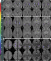
Probabilistic WMI map of 58 very preterm neonates overlaid on (A) the T1-weighted early preterm brain template and (B) a T2-weighted image. Noncystic WMI that occurred at a homologous region in 2 or more very preterm neonates are displayed. WMI seen only in a single neonate are omitted. (A) The cumulative (summed) WMI map of 58 neonates is overlaid on the neonatal brain template in coronal (top) and axial (bottom) planes. The axial planes along the superior to inferior direction are displayed from left to right in the figure. The color bar on the left indicates the color coding of the WMI summation. The maximum value on the map is 14. The cross on the coronal and axial planes of the T1-weighted brain template in each column with (top) and without (bottom) the WMI map is placed at the exactly identical location. (B) The probabilistic WMI map nonlinearly transformed and overlaid on the high-resolution T2-weighted image of a very preterm neonate (postmenstrual age [PMA] 29.7 weeks). First row: the probabilistic WMI map of 58 very preterm neonates nonlinearly transformed and overlaid on the T2-weighted image. Second row: the T2-weighted image. The lesions are seen in the subventricular zone and intermediate zone. The cross on the axial plane of the T2-weighted image in each column with (top) and without (bottom) the WMI map is placed at the same location.
Figure 3
Figure 3. Motor, cognitive, and language outcome-based probabilistic white matter injury (WMI) maps
Probabilistic noncystic WMI map of 58 very preterm neonates based on the motor, cognitive, and language composite scores of Bayley III at 18 months of corrected age. Voxels in blue: WMI that are unique to the neonates who had typical outcomes (scores ≥85); voxels in magenta: WMI that are unique to the neonates who had adverse outcomes (scores <85); voxels in white: WMI common to neonates in both typical and adverse outcome groups. Left pane: the cross is at the territory of the left primary motor area in the neonatal template. Right pane: The cross is at territory of the right inferior parietal lobule in the brain template. The cross indicates the intersecting point for the coronal (top), axial (middle), and sagittal (bottom) planes on the left and the right panes.
Figure 4
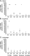
Figure 4. Lobar regional white matter injury (WMI) volume difference and neurodevelopmental outcomes
Regional differences between noncystic WMI volumes in the 4 brain lobes (frontal, parietal, temporal, and occipital) in relation to neurodevelopmental outcomes at 18 months ([A] motor; [B] cognitive; [C] language; Bayley III <85: adverse outcome; Bayley III ≥85: typical outcome). The symbols reflect estimated marginal means of the volumes. Error bars are the 95% confidence intervals. ***p < 0.001; **p < 0.01; *p < 0.05.
Figure 5
Figure 5. Odds ratio (OR) maps of white matter injury (WMI) for adverse motor, cognitive, and language outcomes
OR maps of noncystic WMI for motor (second column), cognitive (third column), and language (fourth column) outcomes overlaid on the T1-weighted neonatal brain template. The first column shows the spatial cumulative WMI map including all noncystic WMI seen in any of the 58 very preterm neonates. Note the different scaling of the images in each column as indicated by each color bar. The purple cross is at exactly the same location in each of the 4 maps. The area with high WMI occurrence rate does not necessarily indicate high risk in developing adverse outcomes. The maximum OR values on the motor, cognitive, and language OR maps are 63.8, 78.9, and 17.5, respectively.

Comment in

Similar articles

Cited by

References

    1. Saigal S, Doyle LW. An overview of mortality and sequelae of preterm birth from infancy to adulthood. Lancet 2008;371:261–269. - PubMed
    1. Back SA, Miller SP. Brain injury in premature neonates: a primary cerebral dysmaturation disorder? Ann Neurol 2014;75:469–486. - PMC - PubMed
    1. Glass HC, Bonifacio SL, Chau V, et al. . Recurrent postnatal infections are associated with progressive white matter injury in premature infants. Pediatrics 2008;122:299–305. - PubMed
    1. Raybaud C, Ahmad T, Rastegar N, Shroff M, Al Nassar M. The premature brain: developmental and lesional anatomy. Neuroradiology 2013;55(suppl 2):23–40. - PubMed
    1. Adams E, Chau V, Poskitt KJ, Grunau RE, Synnes A, Miller SP. Tractography-based quantitation of corticospinal tract development in premature newborns. J Pediatr 2010;156:882–888. - PMC - PubMed

MeSH terms