. 2017 Mar 3;292(9):3827-3840.
doi: 10.1074/jbc.M116.766469. Epub 2017 Jan 18.

Control of Amino Acid Homeostasis by a Ubiquitin Ligase-Coactivator Protein Complex

Affiliations

Control of Amino Acid Homeostasis by a Ubiquitin Ligase-Coactivator Protein Complex

Damian Guerra et al. J Biol Chem. .

Abstract

Intercellular amino acid transport is essential for the growth of all multicellular organisms, and its dysregulation is implicated in developmental disorders. By an unknown mechanism, amino acid efflux is stimulated in plants by overexpression of a membrane-localized protein (GLUTAMINE DUMPER 1 (GDU1)) that requires a ubiquitin ligase (LOSS OF GDU 2 (LOG2). Here we further explore the physiological consequences of the interaction between these two proteins. LOG2 ubiquitin ligase activity is necessary for GDU1-dependent tolerance to exogenous amino acids, and LOG2 self-ubiquitination was markedly stimulated by the GDU1 cytosolic domain, suggesting that GDU1 functions as an adaptor or coactivator of amino acid exporter(s). However, other consequences more typical of a ligase-substrate relationship are observed: disruption of the LOG2 gene increased the in vivo half-life of GDU1, mass spectrometry confirmed that LOG2 ubiquitinates GDU1 at cytosolic lysines, and GDU1 protein levels decreased upon co-expression with active, but not enzymatically inactive LOG2. Altogether these data indicate LOG2 negatively regulates GDU1 protein accumulation by a mechanism dependent upon cytosolic GDU1 lysines. Although GDU1-lysine substituted protein exhibited diminished in vivo ubiquitination, overexpression of GDU1 lysine mutants still conferred amino acid tolerance in a LOG2-dependent manner, consistent with GDU1 being both a substrate and facilitator of LOG2 function. From these data, we offer a model in which GDU1 activates LOG2 to stimulate amino acid export, a process that could be negatively regulated by GDU1 ubiquitination and LOG2 self-ubiquitination.

Keywords: E3 ubiquitin ligase; Loss of GDU1 2; amino acid resistance; amino acid transport; glutamine dumper; protein degradation; ubiquitin; ubiquitin ligase; ubiquitylation (ubiquitination).


Conflict of interest statement

The authors declare that they have no conflicts of interest with the contents of this article

Figures

FIGURE 1.
FIGURE 1.
Wild-type and myristoylation-defective LOG2 proteins restore the Gdu1D amino acid resistance phenotype in GDU1-myc log2-2 plants, but a catalytically inactive LOG2 protein does not. A, 3-week GDU1-myc log2-2 soil-grown plants are larger than age-matched GDU1-myc LOG2 siblings. B, GDU1-myc log2-2, GDU1-myc LOG2, and the non-GDU1-myc lines: control wild-type LOG2 (Col-0) or log2-2 alone seed were plated on GM (right) or GM supplemented with 10 mm phenylalanine (Phe) or leucine (Leu) and photographed after 2 weeks of growth. C, LOG2CCAA-HA, LOG2G2A-HA, and wild-type LOG2-HA transgenes were introduced into the GDU1-myc log2-2 background, triple homozygous seed was plated on GM (filled) or Leu-supplemented GM (open), and the number of green seedlings with expanded true leaves after 10 days of growth at RT were counted. 20 seeds per line were plated in 3 independent experiments. Asterisks indicate significantly different numbers of green seedlings compared with the progenitor GDU1-myc log2-2 line on GM + Leu as assessed by one-way analysis of variance (p < 0.05); all others on GM + Leu were not significantly different from the progenitor. Error bars represent twice the S.E. Seed from GDU1-myc LOG2 (left) and the GDU1-myc log2-2 progenitor (second from left) served as controls. D--F, immunoblots detecting wild-type and mutant LOG2-HA (top panels, anti-HA) and GDU1-myc (middle panels, anti-Myc) protein levels in 10-day-old seedlings grown on GM. Ponc. S (bottom panels), Ponceau stain loading controls. Molecular weight markers in kDa are shown on the left of each blot. Col is wild-type non-transformed control (name of ecotype for Columbia). Genetic background is indicated with bars above the blots. D, protein in LOG2CCAA lines. E, wild-type LOG2 lines. F, LOG2G2A lines. Immunoblots for LOG2CCAA and LOG2G2A lines (D and F) represent signal from 150 μg of total protein. Visualization of LOG2-HA signal (E) required a more sensitive ECL detection kit, longer exposure times, and more total protein compared with D and F (200 or 300 μg for the left and right blot, respectively). White lines in D and F denote removal of uninformative lanes from the same blot.
FIGURE 2.
FIGURE 2.
GDU1-myc steady state levels are higher and degradation is slowed in plants lacking LOG2. A, immunoblot of total protein extracts from GDU1-myc LOG2 (left) and GDU1-myc log2-2 (right) seedlings. Shown is Ponceau S staining for the total protein; molecular weight markers in kDa are on the left. B, semi-log plot of GDU1-myc immunoreactivity in total extracts as a function of time after the addition of the protein synthesis inhibitor CHX to 7-day seedlings from 7 independent experiments. Representative Western blots are shown in the upper right. Linear regressions were drawn in Excel and evaluated by a one-way ANCOVA (supplemental Table 1).
FIGURE 3.
FIGURE 3.
Genetically encoded ubiquitination failed to bypass a requirement for the E3 LOG2 in conferring amino acid resistance. A, two independent GDU1-HA-Ub lines (1 and 2) were crossed to log2-2, and analysis was performed with homozygous GDU1-HA-Ub LOG2 and GDU1-HA-Ub log2-2 F3 siblings. Shown is Western blot of ∼30 μg of total protein from 7-day-old seedlings separated by SDS-PAGE and probed with anti-HA antibodies. The arrow denotes unmodified GDU1-HA-Ub. Top panel, equal exposure indicating different levels of expression between the two independent lines; middle panels, separate exposures for each line for optimal visualization; bottom panel, Ponceau (Ponc. S) loading control. B, same assay as described in Fig. 1C, with the GDU1-HA-Ub expressing transgenic lines in LOG2 and log2-2 backgrounds. Wild-type (Col) without the GDU1-HA-Ub transgene served as the controls. Analysis was conducted as in Fig. 1C.
FIGURE 4.
FIGURE 4.
GDU1 is ubiquitinated at lysines in vitro and in planta. A, in vitro ubiquitination of HA-cGDU1-His6 (wild-type or the K0 mutant lacking all cytosolic lysines) by V5-tagged LOG2. − lanes contain all ubiquitination assay components except LOG2. B, nickel-NTA affinity purification of His6-HA3-GDU1 from transiently transformed N. benthamiana leaves co-infiltrated with LOG2-HA. Input, cell-free supernatant after tissue lysis. NB, not bound (protein that did not bind nickel beads). Eluate, protein eluted from nickel-NTA beads with 350 mm imidazole and heat. The red box in B refers to the region on the corresponding SDS-polyacrylamide gels subjected to in-gel trypsinization and subsequent electrospray ionization-MS/MS tandem mass spectrometry to identify ubiquitination sites. Arrows in A and B denote un-modified HA-cGDU1-His6 and His6-HA3-GDU1, respectively. Numbers to the left of Western blots indicate protein mass in kDa. C, GDU1 amino acid sequence with mass spectrometry-identified ubiquitination sites indicated with underlines (identified in in vitro assays only) or bold underlines (identified in both in vitro and in planta assays). Red text demarcates the cGDU1 region in the GDU1 protein sequence. Serine 127 (underlined) was identified as the sole in vitro ubiquitination site in HA-cGDU1K0-His6. D and E, mass spectra for the parent ion (D) and MS/MS fragmentation (E) indicating in vitro ubiquitination of Lys-148 (ubiquitinated peptides in green).
FIGURE 5.
FIGURE 5.
GDU1 lysines and serine 127 are not required for amino acid resistance. A–C, 7-day-old seedlings expressing different GDU1 proteins with lysine and/or serine 127 substitutions to arginine and alanine, respectively, were grown on GM alone (A) or GM supplemented with 5 mm leucine (B) or 7.5 mm leucine (C) to test amino acid tolerance. The experiment was performed as described in Fig. 1C, except here is expressed as the percentage of green seedlings from 5–7 plantings of ∼25 seedlings from 2 representative independent experiments. Error bars denote twice the S.E. Numbers directly below the graphs denote transgenic line number. GDU1-myc and Col-0 seed served as positive and negative controls, respectively. Italicized abbreviations refer to plants that express the following GDU1-HA transgenes. WT, wild-type GDU1-HA. K9, GDU1-HA in which all lysine codons have been mutated to arginine codons except lysine 9. S127A, GDU1-HA in which the serine 127 codon has been mutated encode alanine. K9 S127A, GDU1-HA in which all lysine codons have been mutated to arginine codons and the serine 127 codon has been mutated to encode alanine. K0 S127A, GDU1-HA in which all lysine codons have been mutated to arginine codons and the serine 127 codon has been mutated encode alanine. D, effect of the lysine/serine substitutions on GDU1 electrophoretic behavior. Extracts of T3 seedlings (50 μg of protein) expressing the indicated GDU1-HA proteins in the wild-type background, visualized with an anti-HA antibody. The thin white line between lanes 1 and 2 represents removal of a non-informative band. Short exposure (right panel) is a shorter exposure of lanes 2 and 3 to demonstrate the difference in laddering between GDU1K9-HA and GDU1S127A-HA.
FIGURE 6.
FIGURE 6.
Substitution of GDU1 lysines did not affect amino acid resistance or its dependence on LOG2. A, germination percentages of F3 seed homozygous for various HA-tagged GDU1 variants on GM supplemented with 7.5 mm leucine in either the homozygous LOG2 (wild-type) or log2-2 background. GDU1 protein designations are as in Fig. 5A. The graph represents three biological replicates (each replicate is a percent germination from a plating of 20–40 seedlings with 1–3 technical replicates) of progeny of double homozygous F2 siblings that express the same GDU1-HA transgene in either the wild-type (LOG2) or the log2-2 background. B, representative plates (top) and legend (bottom) used to generate the data in A. C, GDU1K0 S127A-HA expression levels in 7 independent transgenic lines (4 in the wild-type (LOG2) and 3 in the log2-2 backgrounds) from 10 μg total protein from 14-day-old seedlings visualized with anti-HA antibody. The thin white line between lanes 3 and 4 indicates removal of a non-informative lane; all samples are from the same gel and exposure. D, germination percentage of multiple independent lines (shown in C) expressing GDU1K0 S127A-HA in the wild-type (LOG2) or log2-2 background on 7.5 mm leucine-supplemented media (left). Data represent five replicates, each replicate consisting of 20–50 seed (right). E, germination rates on GM for plant lines in A and D. F, co-expression of wild-type or mutant GDU1-HAs and wild-type or enzymatically inactive (CCAA) LOG2 in N. benthamiana leaves. G, densitometry of immunoreactivities in F. Intensities of wild-type and mutant GDU1-HA bands were quantified with a CCD camera and normalized to the average HA immunoreactivity for each blot. Data are representative of three infiltrations. Error bars correspond to twice the S.E.
FIGURE 7.
FIGURE 7.
LOG2 ubiquitinates itself and GDU1 by different mechanisms, and GDU1 stimulates LOG2 activity in vitro. A, immunoblots of cGDU1 (top, anti-HA) or LOG2 (bottom, anti-LOG2) from ubiquitination assays of HA-cGDU1 and HA-cGDU1G100R (encoded by the log1-1 allele) by LOG2-V5 in the presence of wild-type ubiquitin (WT) or methylated ubiquitin (Me) lacking acceptor lysines for ubiquitin chain formation. Small arrows and arrowheads in A and B indicate mono- and di-ubiquitinated cGDU1, respectively. B, ubiquitination of HA-cGDU1 and HA-cGDU1K0 (lysine-less) by LOG2-V5 in the presence of wild-type ubiquitin (WT) or methylated ubiquitin (Me) and immunoblotted for cGDU1 or LOG2 as in A (top and middle panels). The same reactions were immunoblotted for ubiquitin (bottom panel, anti-ubiquitin antibody). The asterisk indicates monoubiquitinated LOG2. C, LOG2 E3 ligase activity is stimulated by the binding of cGDU1. The plus sign indicates inclusion of E1, E2, Mg-ATP, and Ub. WT, FLAG-cGDU1; log1-1, FLAG-cGDU1G100R. D, time course of cGDU1-activated LOG2 ubiquitination activity. BSA, reaction contained 1 mg·ml−1 bovine serum albumin. E, LOG2 self-ubiquitination occurs in cis but not in trans. Assays contained both wild-type and enzymatically inactive (CCAA) LOG2 (with either a GST or a -V5 tag to allow for adequate electrophoretic separation of the two forms of LOG2) in the presence of HA-cGDU1 (wild-type or the G100R mutant). For all immunoblots, an aliquot of the reaction mixture was fractionated by SDS-PAGE, and proteins were visualized by immunoblotting with the indicated antibody. In all instances, higher migrating forms of the active ligase (but not the inactive ligase) can be observed, indicating LOG2 can promote intramolecular self-ubiquitination (cis-ubiquitination) but not intermolecular (trans-) self-ubiquitination. Single and double asterisks highlight monoubiquitinated LOG2-V5 and GST-LOG2, respectively.
FIGURE 8.
FIGURE 8.
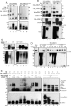
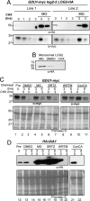
LOG2 and GDU1 are degraded by different proteolytic pathways in vivo. A, LOG2-HA expressed in GDU1-myc log2-2 LOG2-HA complementation lines (visualized from total lysates from seedlings after 7 days in liquid GM media by immunoblotting) after treatment with protein synthesis inhibitor CHX or with a 2-h pretreatment with 100 μm MG132. B, microsomal LOG2-HA from 7-day-old seedlings stable transgenic Arabidopsis were treated with 100 μm MG132 or 1% (v/v) DMSO for 4 h or 200 μg/ml CHX for 1 h before microsomal purification. Equal total microsomal protein was fractionated by SDS-PAGE, and LOG2-HA was visualized with anti-HA antibodies. C, 7-day-old seedlings grown in liquid GM media were treated with solvent control (DMSO), proteasome inhibitors (MG132 (MG) or bortezomib IBRTZ)), wortmannin (WRTM, a vacuolar maturation inhibitor), or concanamycin A (ConCA, which inhibits vacuolar acidification) for 3 h before the addition of CHX. D, same as C with seedlings that express HA-IAA1, an HA-tagged auxin response transcriptional repressor IAA1, a known proteasomal target. Equal total protein was loaded, and immunoblot analysis was performed using anti-myc (for GDU1) or anti-HA (for HA-IAA1). Ponceau S staining indicates total protein.
FIGURE 9.
FIGURE 9.
A model for the dependence of the Gdu1D phenotype on LOG2 ubiquitin ligase activity. In the presence of LOG2 (yellow), GDU1 (green) overexpression activates an amino acid transport system either by directly associating with an amino acid exporter (red), by functioning through as yet-undiscovered facilitator(s) (X), or by down-regulating an inhibitor of the transporter. This process depends in part on GDU1-activated LOG2 ubiquitination activity. GDU1 may also be an adaptor protein that aids in the recognition of amino acid exporters or unknown inhibitors/facilitators. P. M., plasma membrane.

Similar articles

Cited by

References

    1. Pickart C. M., and Eddins M. J. (2004) Ubiquitin: structures, functions, mechanisms. Biochim. Biophys. Acta 1695, 55–72 - PubMed
    1. Lucero P., Peñalver E., Vela L., and Lagunas R. (2000) Monoubiquitination is sufficient to signal internalization of the maltose transporter in Saccharomyces cerevisiae. J. Bacteriol 182, 241–243 - PMC - PubMed
    1. Uhrig R. G., She Y.-M., Leach C. A., and Plaxton W. C. (2008) Regulatory monoubiquitination of phosphoenolpyruvate carboxylase in germinating castor oil seeds. J. Biol. Chem. 283, 29650–29657 - PMC - PubMed
    1. Guerra D. D., and Callis J. (2012) Ubiquitin on the Move: the ubiquitin modification pathway plays diverse roles in the regulation of Er-and membrane-localized proteins. Plant Physiol. 160, 56–64 - PMC - PubMed
    1. Kerscher O., Felberbaum R., and Hochstrasser M. (2006) Modification of proteins by ubiquitin and ubiquitin-like proteins. Annu. Rev. Cell Dev. Biol. 22, 159–180 - PubMed

Publication types