. 2017 May 5;292(18):7566-7577.
doi: 10.1074/jbc.M116.760710. Epub 2017 Jan 18.

A novel microRNA located in the TrkC gene regulates the Wnt signaling pathway and is differentially expressed in colorectal cancer specimens

Affiliations

A novel microRNA located in the TrkC gene regulates the Wnt signaling pathway and is differentially expressed in colorectal cancer specimens

Sadat Dokanehiifard et al. J Biol Chem. .

Abstract

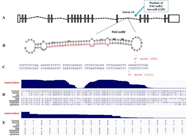
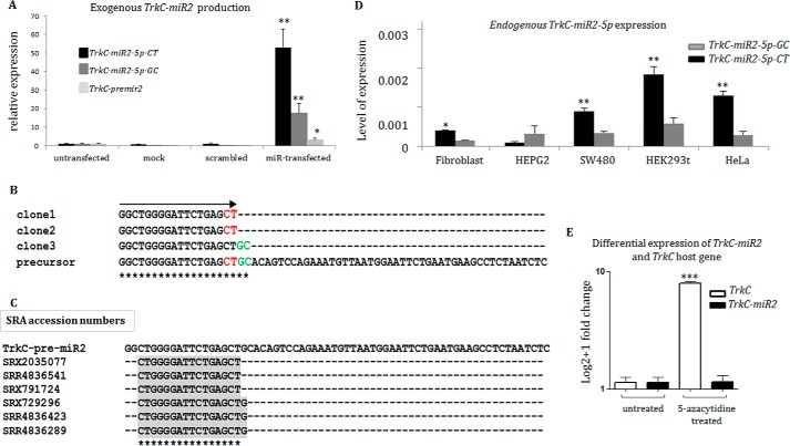
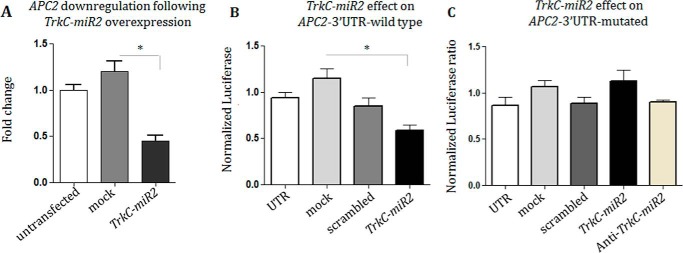
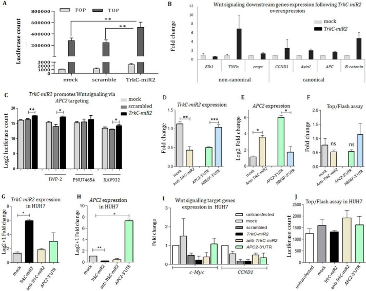
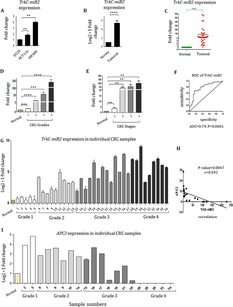
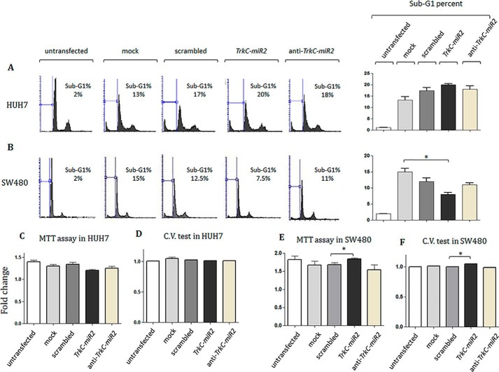
Tropomyosin receptor kinase C (TrkC) is involved in cell survival, apoptosis, differentiation, and tumorigenesis. TrkC diverse functions might be attributed to the hypothetical non-coding RNAs embedded within the gene. Using bioinformatics approaches, a novel microRNA named TrkC-miR2 was predicted within the TrkC gene capable of regulating the Wnt pathway. For experimental verification of this microRNA, the predicted TrkC-premir2 sequence was overexpressed in SW480 cells, which led to the detection of two mature TrkC-miR2 isomiRs, and their endogenous forms were detected in human cell lines as well. Later, an independent promoter was deduced for TrkC-miR2 after the treatment of HCT116 cells with 5-azacytidine, which resulted in differential expression of TrkC-miR2 and TrkC host gene. RT-quantitative PCR and luciferase assays indicated that the APC2 gene is targeted by TrkC-miR2, and Wnt signaling is up-regulated. Also, Wnt inhibition by using small molecules along with TrkC-miR2 overexpression and TOP/FOP flash assays confirmed the positive effect of TrkC-miR2 on the Wnt pathway. Consistently, TrkC-miR2 overexpression promoted SW480 cell survival, which was detected by flow cytometry, MTT (3-(4,5-dimethylthiazol-2-yl)-2,5-diphenyltetrazolium bromide) assays, and crystal violate analysis. RT-qPCR analysis revealed that TrkC-miR2 is significantly up-regulated (∼70 times) in colorectal tumor tissues compared with their normal pairs. Moreover, the TrkC-miR2 expression level discriminated grades of tumor malignancies, which was consistent with its endogenous levels in HCT116, HT29, and SW480 colorectal cancer cell lines. Finally, an opposite expression pattern was observed for TrkC-miR2 and the APC2 gene in colorectal cancer specimens. In conclusion, here we introduce TrkC-miR2 as a novel regulator of Wnt signaling, which might be a candidate oncogenic colorectal cancer biomarker.

Keywords: Biomarker; Colorectal cancer; Survival; TrkC-miR2; Wnt signaling; annexin; biomarker; cancer; cell cycle; colorectal cancer.


Conflict of interest statement

The authors declare that they have no conflicts of interest with the contents of this article

Figures

Figure 1.
Figure 1.
Bioinformatics prediction of TrkC-miR2 encoded within the 14th intron of human TrkC gene. A, TrkC gene introns (broken lines) and exons (rectangles) adapted from Ensemble are shown. The position of the stem loop encoding TrkC-miR2 is shown at in the 17-kb vicinity of the newly discovered hsa-miR-11181. B, predicted stem loop encoding TrkC-miR2. The red sequence was predicted by SSC profiler as the TrkC-miR2 mature form. C, Drosha enzyme cutting sites, predicted by Microprocessor SVM tool, are shown on the sequence of hairpin structure. Blat search by the UCSC genome browser shows high conservation of TrkC-premir2 (D) and TrkC-miR2–5p (E) between several organisms.
Figure 2.
Figure 2.
TrkC-miR2–5p sequence characterization and evidence for an independent promoter. A, overexpression of TrkC-premir2 in SW480 cells and detection of predicted TrkC-miR2–5p mature forms (-CT and -GC isomiRs) by RT-qPCR. Note that TrkC-miR2–5p-CT isomiR is dominantly produced in the transfected cells. B, sequencing results of three TA vector clones containing TrkC-miR2–5p sequences created in A. Clones 1 and 2 show the sequence of TrkC-miR2–5p-CT isomiR, and clone 3 shows the sequence of TrkC-miR2–5p-GC isomiR. TrkC-miR2 sequences are aligned with the sequence of their precursor. The significance of asterisks in panels C and B is the complete matching of aligned sequences at each position. C, shown is the presence of TrkC-miR2 in the SRA data. Three reads for detected TrkC-miR2-5p-CT and TrkC-miR2-5p-CTG isomiRs are shown. However, no read for TrkC-miR2-5p-GC was detected in the SRA data set. D, shown is the expression level of TrkC-miR2–5p-CT and -GC isomiRs in several human cell lines. TrkC-miR2–5p-CT relative expression was higher than TrkC-miR2–5p-GC isomiR in most of these cell lines. E, treatment of HCT-116 cells with epidrug resulted in TrkC expression elevation (100 times). However, it did not have such an effect on TrkC-miR2–5p expression level. Error bars indicate S.D. of duplicate experiments. U48 RNA and GAPDH were used as internal controls for the amplifications. *, p < 0.05; **, p < 0.01; ***, p < 0.001.
Figure 3.
Figure 3.
TrkC-miR2 direct interaction with its predicted target gene. A, RT-qPCR result shows APC2 gene down-regulation after TrkC-premir2 overexpression in the SW480 cell line compared with the related controls. B, dual luciferase assay supported TrkC-miR2 direct interaction with 3′-UTR sequence of APC2 target gene. C, lack of interaction between the overexpressed TrkC-miR2 and mutated APC2,3′-UTR detected by dual luciferase assay and supported direct interaction between this miRNA and the wild-type sequence of APC2,3′-UTR sequence shown in B. *, p < 0.05.
Figure 4.
Figure 4.
TrkC-miR2 expression alteration effect on the Wnt signaling pathway. A, Wnt signaling up-regulation after TrkC-miR2 overexpression in the SW480 cells. Luciferase activity was significantly increased in the cells overexpressing TrkC-premir2 compared with the controls. B, increased expression level of the genes involved in both canonical and non-canonical Wnt signaling pathway after TrkC-miR2 overexpression. C, Wnt inhibitor small molecules and TrkC-miR2 co-treatment effect on the Wnt signaling is shown through Top/flash assay. Although Wnt signaling is down-regulated after the XAV932 and IWP-2 small molecules application, this down-regulation has been compensated by TrkC-miR2 overexpression compared with the controls. When the cells were treated with the PNU74654 small molecule, significant Wnt signaling alteration was not detected with or without TrkC-miR2 overexpression. Error bars indicate S.D. of three experiments. D, successful down-regulation of TrkC-miR2 using anti-TrkC-miR2 sequence in SW480 cells (compared with the mock control). Also, APC2-3′-UTR overexpression as a scavenger, resulted in down-regulation of TrkC-miR2 (compared with the non-scavenger HBEGF-3′-UTR sequence), detected by RT-qPCR. Expression data were normalized against U48 as an internal control. E, RT-qPCR results showed significant up-regulation of APC2 expression after the down-regulation of TrkC-miR2 (using anti-TrkC-miR2) or after the scavenging of TrkC-miR2 (by APC2-3′-UTR overexpression) in SW480 cells. Expression data were normalized against GAPDH as an internal control. F, top/Fop flash assay after transfection of anti-TrkC-miR2 in SW480 cells showing non-significant Wnt signaling reduction compared with the mock transfected control. Also, overexpression of APC2-3′-UTR as a TrkC-miR2 scavenger again resulted in non-significant Wnt signaling attenuation compared with the off-target HBEGF-3′-UTR overexpression. ns, not significant. G, RT-qPCR analysis of TrkC-miR2 expression in HUH7 cell lines after TrkC-premiR2, anti-TrkC-miR2, and APC2-3′-UTR overexpression compared with mock control. TrkC-miR2 level was significantly increased in the cells overexpressing TrkC-premiR2; however, anti-TrkC-miR2 and APC2-3′-UTR scavenger constructs were not capable of significant reduction in TrkC-miR2 level. H, shows APC2 expression alteration after TrkC-premiR2, anti-TrkC-miR2, and APC2-3′-UTR overexpression. APC2-3′-UTR-specific primers were used for detection of APC2 expression alteration. Only successful overexpression of TrkC-miR2 (G) has resulted in significant reduction of APC2 transcripts. I, RT-qPCR against c-Myc and CCND1 genes (as the Wnt signaling target genes) after the overexpression of interested constructs. Data indicate no significant Wnt signaling alteration after the TrkC-miR2 expression alteration in Huh7 cells. J, shown is the Top/flash assay in the HUH7 cell line before and after TrkC-miR2 expression alteration. Similar to I, no significant Wnt signaling alteration was detected. *, p < 0.05; **, p < 0.01; ***, p < 0.001.
Figure 5.
Figure 5.
Implication of TrkC-miR2 in colorectal cancer. A, TrkC-miR2-5p-CT expression level in HCT-116 and SW480 cells was 2- and 4-fold higher than HT29 cell line (p < 0. 005), respectively. B, TrkC-miR2 expression status in 36 CRC tumor (T) tissues and normal (N) pairs. C, Mann-Whitney analysis indicated that TrkC-miR2 has been significantly increased in CRCs (∼70-fold) compared with the paired adjacent non-CRC tissue samples (p < 0.0001). D, increased expression of TrkC-miR2 detected in the more advanced grades of CRCs compared with the normal ones (p < 0.001). E, TrkC-miR2 expression in different stages of CRC samples. Error bars indicate S.E. of tetraplicate experiments. F, ROC curve analysis of TrkC-miR2 expression in CRC patients. The up-regulated TrkC-miR2 expression yielded an area under the curve (AUC) value of 0.79 (95% confidence interval: 0.6714–0.8944) with 75% sensitivity and 75% specificity, supported TrkC-miR2 expression as a diagnostic value for discriminating CRC from healthy controls. G, TrkC-miR2 expression analysis in individual samples, distributed in high and low grade samples. Samples are shown by the numbers. H, a significant negative correlation was calculated between TrkC-miR2 and APC2 expression with a correlation coefficient (r) of −0.592 and a significant p value of 0.0046. I, APC2 expression analysis in individual high and low grade samples. Error bars indicate S.D. of duplicated experiments. *, p < 0.05; **, p < 0.01; ***, p < 0.001; ****, p < 0.0001.
Figure 6.
Figure 6.
Cell cycle and survival analysis for the cells overexpressing TrkC-premir2. A and B, shown is flow cytometry propidium iodide staining 36 h after transfection of two different cell lines in which TrkC-premir2 has been overexpressed or down-regulated. Although no significant alteration was detected for HUH7 cell line (A), t test analysis indicated a significant reduction in sub-G1 cell cycle distribution of SW480 (B)-transfected cells after TrkC-miR2 overexpression. However, knockdown of TrkC-miR2 in both cell types did not significantly change the sub-G1 percentages of the cells. C and D, MTT and crystal violet assay against HUH7 cells under TrkC-miR2 expression alteration. No significant cell survival alteration rate was calculated in these situations. E and F, MTT and crystal violet assay against SW480 cells under TrkC-miR2 expression alteration. A significant cell survival alteration rate was calculated when TrkC-miR2 was overexpressed. *, p < 0.05

Similar articles

Cited by

References

    1. Reichardt L. F. (2006) Neurotrophin-regulated signalling pathways. Philos. Trans. R. Soc. Lond. B. Biol. Sci. 361, 1545–1564 - PMC - PubMed
    1. McGregor L. M., McCune B. K., Graff J. R., McDowell P. R., Romans K. E., Yancopoulos G. D., Ball D. W., Baylin S. B., and Nelkin B. D. (1999) Roles of trk family neurotrophin receptors in medullary thyroid carcinoma development and progression. Proc. Natl. Acad. Sci. U.S.A. 96, 4540–4545 - PMC - PubMed
    1. Genevois A.-L., Ichim G., Coissieux M.-M., Lambert M.-P., Lavial F., Goldschneider D., Jarrosson-Wuilleme L., Lepinasse F., Gouysse G., Herceg Z., Scoazec J. Y., Tauszig-Delamasure S., and Mehlen P. (2013) Dependence receptor TrkC is a putative colon cancer tumor suppressor. Proc. Natl. Acad. Sci. U.S.A. 110, 3017–3022 - PMC - PubMed
    1. Jin W., Kim G. M., Kim M. S., Lim M. H., Yun C., Jeong J., Nam J.-S., and Kim S.-J. (2010) TrkC plays an essential role in breast tumor growth and metastasis. Carcinogenesis 31, 1939–1947 - PubMed
    1. Luo Y., Kaz A. M., Kanngurn S., Welsch P., Morris S. M., Wang J., Lutterbaugh J. D., Markowitz S. D., and Grady W. M. (2013) NTRK3 is a potential tumor suppressor gene commonly inactivated by epigenetic mechanisms in colorectal cancer. PLoS Genet. 9, e1003552. - PMC - PubMed

Publication types