

A Man with Pancreatic Head Mass Lesion on Endoscopic Ultrasound and Granuloma on Cytopathology
- PMID: 28100998
- PMCID: PMC5216211
- DOI: 10.1159/000448875
A Man with Pancreatic Head Mass Lesion on Endoscopic Ultrasound and Granuloma on Cytopathology
Abstract
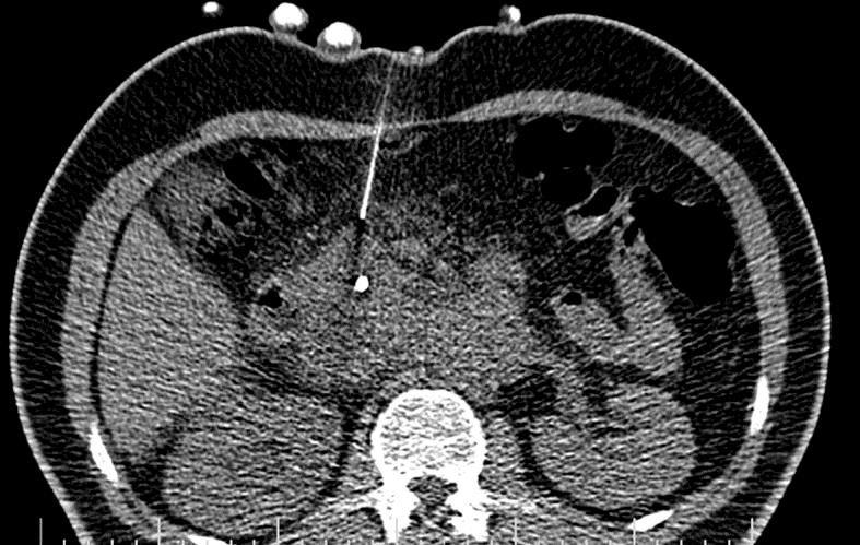
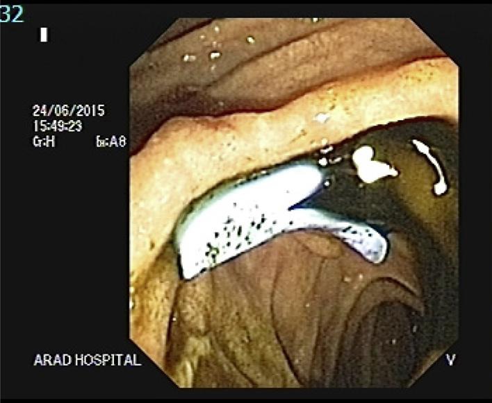
Primary pancreatic lymphoma is an unlikely malignancy accounting for less than 0.5% of pancreatic tumors. Clinical presentation is often nonspecific and may be clinically misdiagnosed as pancreatic adenocarcinoma. Here we present an Iranian case of primary pancreatic lymphoma in a 47-year-old male suffering from jaundice and 20% weight loss. Endoscopic ultrasound revealed a mixed echoic mass lesion at the head of pancreas. The patient underwent endoscopic ultrasound-guided fine needle aspiration of solid pancreatic mass and histopathologic diagnosis revealed granuloma. Computed tomography-guided core needle biopsy was performed and eventually histological examination showed granuloma that was coherent with the diagnosis of primary pancreatic lymphoma. Primary pancreatic lymphoma is a rare entity presenting with nonspecific symptoms, laboratory and radiological findings. Computed tomography results in combination with clinical and radiological studies generally provide guidance for appropriate investigation.
Keywords: Computed tomography-guided core needle biopsy; Endoscopic ultrasound; Endoscopic ultrasound-guided fine needle aspiration; Primary pancreatic lymphoma.
Conflict of interest statement
The authors declare that they have no competing interests.
Figures


References
-
- Orootan FH, Mansour Ghanaie F, Ghofrani H. Primary pancreatic lymphoma: a case report and literature review. Med J Islam Repub Iran. 2001;15:117–121.
Publication types
LinkOut - more resources
Full Text Sources
Other Literature Sources
