

Heads-Up Macular Surgery with a 27-Gauge Microincision Vitrectomy System and Minimal Illumination
- PMID: 28101044
- PMCID: PMC5216224
- DOI: 10.1159/000452993
Heads-Up Macular Surgery with a 27-Gauge Microincision Vitrectomy System and Minimal Illumination
Abstract
Objective: We combined heads-up 3-dimensional (3D) 27-gauge microincision vitrectomy surgery (27GMIVS) with a very low-intensity illumination system.
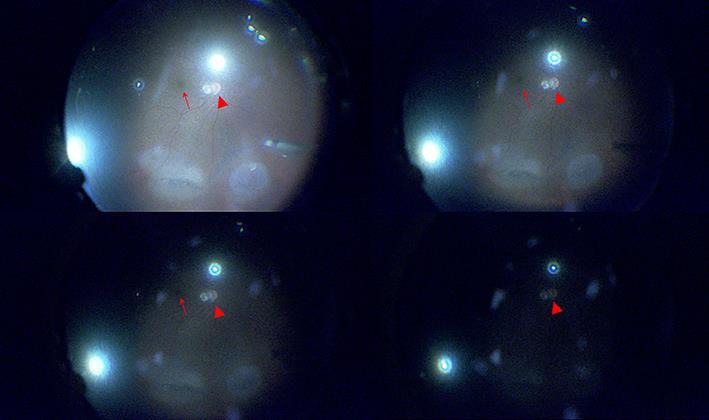
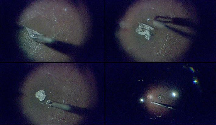
Methods: This study was based on a retrospective, interventional case series of 6 eyes of 6 patients with macular disease. All patients underwent heads-up 3D 27GMIVS and the power of the intraocular illuminator was set to its minimum level, 1% (approximately 0.1 lm), throughout the surgery.
Results: We found that the procedure was easy when the heads-up 3D system was used, but not through the eyepiece of a microscope. All surgeries were successfully finished without any complications. Postoperative visual acuity was restored or maintained in all eyes during the follow-up period.
Conclusion: Heads-up, 3D system-assisted 27GMIVS with minimal illumination enabled excellent intraoperative visualization of retinal tissues, caused minimal phototoxicity to the macular retinal cells, and might therefore represent the next step in the development of an ideal, minimally invasive method of treating macular disease.
Keywords: 27-Gauge vitrectomy; Epiretinal membrane; Heads-up surgery; Macular hole; Phototoxicity.
Conflict of interest statement
The funders had no role in the design or conduct of the study; collection, management, analysis, or interpretation of the data; preparation, review, or approval of the manuscript; or the decision to submit the manuscript for publication.
Figures
References
-
- Michels RG. Vitreous surgery for macular pucker. Am J Ophthalmol. 1981;92:628–639. - PubMed
-
- Oshima Y, Awh CC, Tano Y. Self-retaining 27-gauge transconjunctival chandelier endoillumination for panoramic viewing during vitreous surgery. Am J Ophthalmol. 2007;143:166–167. - PubMed
-
- Yanagi Y, Iriyama A, Jang WD, Kadonosono K. Evaluation of the safety of xenon/bandpass light in vitrectomy using the A2E-laden RPE model. Graefes Arch Clin Exp Ophthalmol. 2007;245:677–681. - PubMed
-
- Eckardt C, Paulo EB. Heads-up surgery for vitreoretinal procedures: an experimental and clinical study. Retina. 2016;36:137–147. - PubMed
Publication types
LinkOut - more resources
Full Text Sources
Other Literature Sources
