. 2017 Jan 4:7:657.
doi: 10.3389/fphys.2016.00657. eCollection 2016.

Elastase-Induced Parenchymal Disruption and Airway Hyper Responsiveness in Mouse Precision Cut Lung Slices: Toward an Ex vivo COPD Model

Affiliations

Elastase-Induced Parenchymal Disruption and Airway Hyper Responsiveness in Mouse Precision Cut Lung Slices: Toward an Ex vivo COPD Model

Eline M Van Dijk et al. Front Physiol. .

Abstract

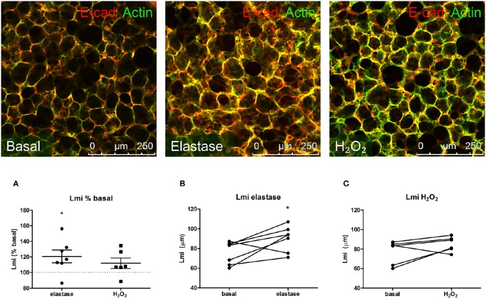
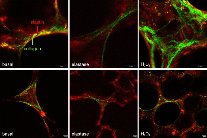
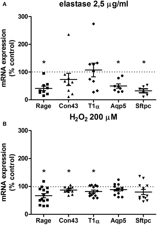
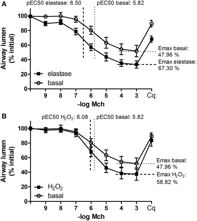
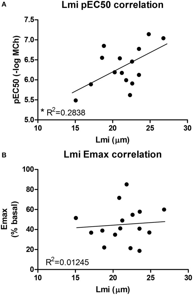
Background: COPD is a progressive lung disease characterized by emphysema and enhanced bronchoconstriction. Current treatments focused on bronchodilation can delay disease progression to some extent, but recovery or normalization of loss of lung function is impossible. Therefore, novel therapeutic targets are needed. The importance of the parenchyma in airway narrowing is increasingly recognized. In COPD, the parenchyma and extracellular matrix are altered, possibly affecting airway mechanics and enhancing bronchoconstriction. Our aim was to set up a comprehensive ex vivo Precision Cut Lung Slice (PCLS) model with a pathophysiology resembling that of COPD and integrate multiple readouts in order to study the relationship between parenchyma, airway functionality, and lung repair processes. Methods: Lungs of C57Bl/6J mice were sliced and treated ex vivo with elastase (2.5 μg/ml) or H2O2 (200 μM) for 16 h. Following treatment, parenchymal structure, airway narrowing, and gene expression levels of alveolar Type I and II cell repair were assessed. Results: Following elastase, but not H2O2 treatment, slices showed a significant increase in mean linear intercept (Lmi), reflective of emphysema. Only elastase-treated slices showed disorganization of elastin and collagen fibers. In addition, elastase treatment lowered both alveolar Type I and II marker expression, whereas H2O2 stimulation lowered alveolar Type I marker expression only. Furthermore, elastase-treated slices showed enhanced methacholine-induced airway narrowing as reflected by increased pEC50 (5.87 at basal vs. 6.50 after elastase treatment) and Emax values (47.96 vs. 67.30%), and impaired chloroquine-induced airway opening. The increase in pEC50 correlated with an increase in mean Lmi. Conclusion: Using this model, we show that structural disruption of elastin fibers leads to impaired alveolar repair, disruption of the parenchymal compartment, and altered airway biomechanics, enhancing airway contraction. This finding may have implications for COPD, as the amount of elastin fiber and parenchymal tissue disruption is associated with disease severity. Therefore, we suggest that PCLS can be used to model certain aspects of COPD pathophysiology and that the parenchymal tissue damage observed in COPD contributes to lung function decline by disrupting airway biomechanics. Targeting the parenchymal compartment may therefore be a promising therapeutic target in the treatment of COPD.

Keywords: airway mechanics; chronic obstructive pulmonary disease; extracellular matrix.


Figures

Figure 1
Figure 1
Elastase, but not H2O2 treatment increases Lmi. PCLS were exposed to elastase (2.5 μg/ml, n = 6) or H2O2 (200 μM, n = 5) for 16 h. After stimulation, slices were washed twice with medium and incubated for 24 h in medium. (A) Following incubation, slices were stained for F-actin filaments (green) and E-cadherin (red) and Lmi was assessed as % basal. (B,C) Lmi following treatments shown in μm. The statistical significance of differences between means was determined on log transformed data by Student's t-test (A) or by Student's t-test (B,C). Data represent mean ± SEM, *p < 0.05 compared to basal control.
Figure 2
Figure 2
Elastase, but not H2O2 treatment disrupts the structural organization of elastin and collagen. PCLS were exposed to elastase (2.5 μg/ml) or H2O2 (200 μM) for 16 h. After stimulation, slices were washed twice with medium and incubated for 24 h in medium. 2-Photon and multiphoton excitation fluorescence (MPEF) imaging were used to visualize collagen and elastin polymers. Following elastase, but not H2O2, elastin and collagen showed a disrupted fiber organization.
Figure 3
Figure 3
Elastase and H2O2 treatment alter mRNA expression levels of alveolar makers. PCLS were exposed to (A) elastase (2.5 μg/ml, n = 9) or (B) H2O2 (200 μM, n = 14) for 16 h. After stimulation, slices were washed twice with medium and incubated for 24 h in medium. The statistical significance of differences between means was determined on log transformed data by Student's t-test followed by a Bonferroni correction. Data represent mean ± SEM, *p < 0.05 compared to basal control.
Figure 4
Figure 4
Elastase, but not H2O2 treatment enhances MCh-induced airway narrowing. PCLS were exposed to elastase (2.5 μg/ml, n = 10) or H2O2 (200 μM, n = 10) for 16 h. After stimulation, slices were washed twice with medium and incubated for 24 h in medium. Following incubation, MCh-induced airway narrowing was assessed. Lung slice images were captured in time-lapse (1 frame per 2 s) using an inverted phase contrast microscope (Eclipse, TS100; Nikon). Airway luminal area was quantified using image acquisition software (NIS-elements; Nikon), and expressed as percent basal. (A) MCh-induced airway narrowing following elastase treatment. Elastase treatment increased pEC50 values significantly (p < 0.05, compared to basal control). (B) MCh-induced airway narrowing following H2O2 treament. The statistical significance of differences between means was determined on log transformed data by one-way ANOVA followed by Bonferonni testing. Data represent mean ± SEM.
Figure 5
Figure 5
An increased pEC50 value correlates with an increased Lmi. (A) Lmi (μm) and pEC50 values for all conditions were combined. It was found that an increased pEC50 value correlates with an increased Lmi (R2 = 0.2838, p < 0.05). (B) Emax values did not correlate with Lmi.
Figure 6
Figure 6
Elastase, but not H2O2 treatment impairs chloroquine-induced airway opening. PCLS were exposed to elastase (2.5 μg/ml, n = 8) or H2O2 (200 μM, n = 8) for 16 h. After stimulation, slices were washed twice with medium and incubated for 24 h in medium. Following incubation, airway narrowing was induced by an increasing dose of MCh. Subsequently, airway relaxation was induced with chloroquine. Elastase, but not H2O2 treatment, impaired chloroquine-induced relaxation of contracted airways. The statistical significance of differences between means was determined on log transformed data by Student's t-test. Data represent mean ± SEM, *p < 0.05 compared to basal control.

Similar articles

Cited by

References

    1. Abraham T., Hogg J. (2010). Extracellular matrix remodeling of lung alveolar walls in three dimensional space identified using second harmonic generation and multiphoton excitation fluorescence. J. Struct. Biol. 171, 189–196. 10.1016/j.jsb.2010.04.006 - DOI - PubMed
    1. An S. S., Bai T. R., Bates J. H., Black J. L., Brown R. H., Brusasco V., et al. . (2007). Airway smooth muscle dynamics: a common pathway of airway obstruction in asthma. Eur. Respir. J. 29, 834–860. 10.1183/09031936.00112606 - DOI - PMC - PubMed
    1. Barnes P. J. (2016). Inflammatory mechanisms in patients with chronic obstructive pulmonary disease. J. Allergy Clin. Immunol. 138, 16–27. 10.1016/j.jaci.2016.05.011 - DOI - PubMed
    1. Barnes P. J., Shapiro S. D., Pauwels R. A. (2003). Chronic obstructive pulmonary disease: molecular and cellular mechanisms. Eur. Respir. J. 22, 672–688. 10.1183/09031936.03.00040703 - DOI - PubMed
    1. Bidan C. M., Veldsink A. C., Meurs H., Gosens R. (2015). Airway and extracellular matrix mechanics in COPD. Front. Physiol. 6:346. 10.3389/fphys.2015.00346 - DOI - PMC - PubMed

LinkOut - more resources