. 2017 Jan 4:7:2058.
doi: 10.3389/fmicb.2016.02058. eCollection 2016.

A Disease Model of Muscle Necrosis Caused by Aeromonas dhakensis Infection in Caenorhabditis elegans

Affiliations

A Disease Model of Muscle Necrosis Caused by Aeromonas dhakensis Infection in Caenorhabditis elegans

Po-Lin Chen et al. Front Microbiol. .

Abstract

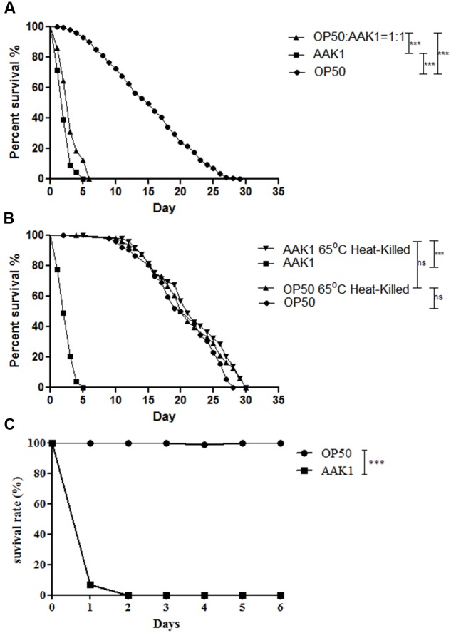
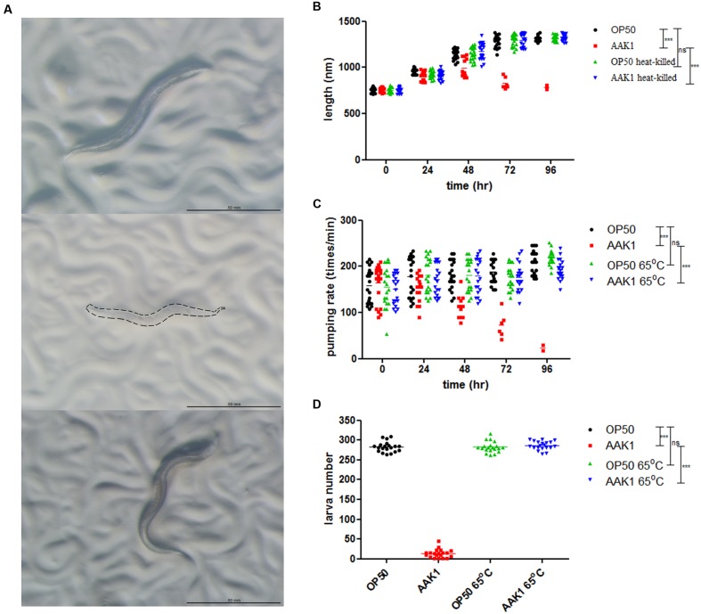
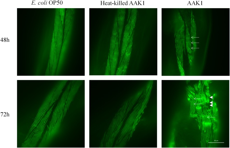
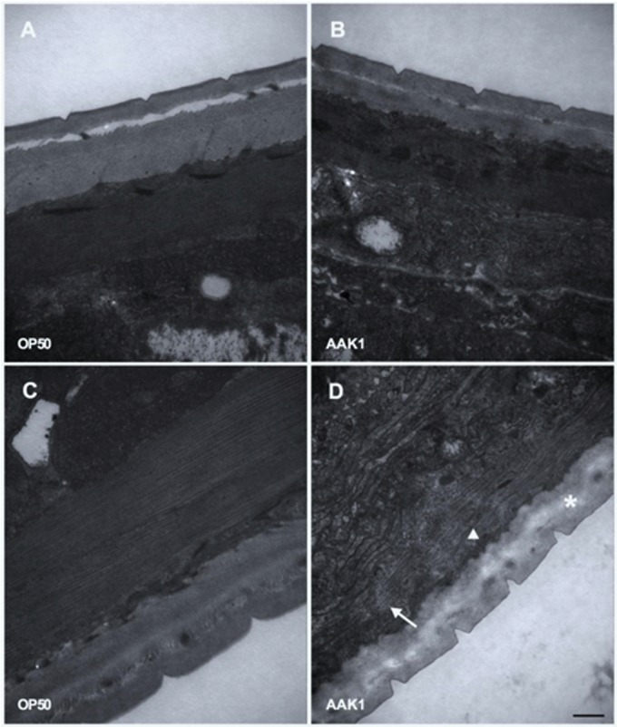
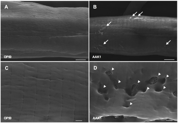
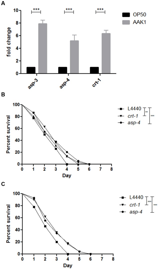
A variety of bacterial infections cause muscle necrosis in humans. Caenorhabditis elegans has epidermis and bands of muscle that resemble soft-tissue structures in mammals and humans. Here, we developed a muscle necrosis model caused by Aeromonas dhakensis infection in C. elegans. Our data showed that A. dhakensis infected and killed C. elegans rapidly. Characteristic muscle damage in C. elegans induced by A. dhakensis was demonstrated in vivo. Relative expression levels of host necrosis-associated genes, asp-3, asp-4, and crt-1 increased significantly after A. dhakensis infection. The RNAi sensitive NL2099 rrf-3 (pk1426) worms with knockdown of necrosis genes of crt-1 and asp-4 by RNAi showed prolonged survival after A. dhakensis infection. Specifically knockdown of crt-1 and asp-4 by RNAi in WM118 worms, which restricted RNAi only to the muscle cells, conferred significant resistance to A. dhakensis infection. In contrast, the severity of muscle damage and toxicity produced by the A. dhakensis hemolysin-deletion mutant is attenuated. In another example, shiga-like toxin-producing enterohemorrhagic E. coli (EHEC) known to elicit toxicity to C. elegans with concomitant enteropathogenicty, did not cause muscle necrosis as A. dhakensis did. Taken together, these results show that Aeromonas infection induces muscle necrosis and rapid death of infected C. elegans, which are similar to muscle necrosis in humans, and then validate the value of the C. elegans model with A. dhakensis infection in studying Aeromonas pathogenicity.

Keywords: Aeromonas dhakensis; Caenorhabditis elegans; disease model; infection; muscle necrosis.


Figures

FIGURE 1
FIGURE 1
Aeromonas dhakensis shortens the life span of Caenorhabditis elegans. (A) Worms infected with A. dhakensis and A. dhakensis mixed with Escherichia coli OP50 (1:1) had a significantly shorter life span when compared with control E. coli OP50. (B) A. dhakensis AAK1 showed toxicity when compared with heat-killed A. dhakensis or control E. coli OP50. (C) In liquid-toxic (LT) assay, the survival rate of C. elegans infected with A. dhakensis was significantly lower than those with control strain E. coli OP50. ∗∗∗P < 0.0001 by the Mantel-Cox log-rank test.
FIGURE 2
FIGURE 2
Aeromonas dhakensis infection induces morphological and physiological changes in C. elegans. (A) Morphology of C. elegans treated with A. dhakensis AAK1 for 72 h (Top to bottom: E. coli OP50, A. dhakensis AAK1, 65°C heat-killed A. dhakensis AAK1). At 72 h, AAK1 infection resulted in total lysis of body. The corpse of AAK1 infected worm was marked by dashed line. The scale bar indicates 500 μm. (B) The body length of N2 C. elegans was significantly decreased after A. dhakensis AAK1 infection when compared with the control E. coli OP50 (P < 0.0001) by two-way ANOVA test. In contrast, 65°C heat-killed bacteria did not affect the size of the animals. The average feeding rate measured by pharyngeal movement (pumping times/min) (C) and brood size (D) of N2 worms infected with AAK1were significantly reduced, as compared with worms fed on OP50, OP50-65°C, or AAK1-65°C, ∗∗∗P < 0.0001.
FIGURE 3
FIGURE 3
Aeromonas dhakensis induces muscle damage in Caenorhabditis elegans. Bending of muscle fiber (arrows) was observed in worms with A. dhakensis AAK1 infection at 48 h. Rupture of muscle fibers (arrowheads) was obvious at 72 h. In contrast, E. coli OP50 or heat-killed A. dhakensis AAK1 did not cause muscle damage. Scale bar indicates 50 μm.
FIGURE 4
FIGURE 4
Transmission electron microscopic analysis. The morphological changes in C. elegans after feeding with A. dhakensis AAK1 (right panels) or E. coli OP50 (left panels) for 48 h (upper panels) or 72 h (lower panels) were observed with transmission electron microscopy (A–D). Patch hypo-dense lesions (arrow), wavy change of myosin filaments (arrowhead), and decreased density of hypodermis (asterisks) were observed at 72 h after AAK1 infection (D). Scale bar indicates 500 nm.
FIGURE 5
FIGURE 5
Scanning electron microscopic analysis. Features of N2 animals infected with A. dhakensis AAK1 (right panels, B,D) and control E. coli OP50 (left panels, A,C) for 48 h (upper panels) and 72 h (lower panels) observed by scanning electron microscopy. At 48 h, AAK1 cells that had adhered to animal surface are indicated by arrows (B). Uneven surface and punctate lesions caused by invasion of AAK1 are indicated by arrowheads at 72 h (D). Scale bars in the upper panel indicate 5 μm, and those in the lower panel indicate 1 μm.
FIGURE 6
FIGURE 6
Aeromonas dhakensis induces necrosis in C. elegans. (A) Relative expression levels of asp-3, asp-4, and crt-1 genes measured by quantitative real-time PCR (qPCR) in C. elegans infected by A. dhakensis AAK1 for 72 h were higher than C. elegans fed by E. coli OP50. (B) The RNAi-sensitive worms, NL2099 rrf-3 (pk1426), with knockdown of crt-1 or asp-4 by RNAi showed significantly enhanced survival after A. dhakensis infection, relative to the control animals with RNAi control (L4440). (C) The WM118 worms with depressed expression of crt-1 or asp-4 by RNAi restricted to the muscle were more resistant to A. dhakensis infection than RNAi control (L4440). ∗∗P < 0.01; ∗∗∗P < 0.0001.

Similar articles

Cited by

References

    1. Anaya D. A., Dellinger E. P. (2007). Necrotizing soft-tissue infection: diagnosis and management. Clin. Infect. Dis. 44 705–710. 10.1086/511638 - DOI - PubMed
    1. Aravena-Roman M., Harnett G. B., Riley T. V., Inglis T. J., Chang B. J. (2011). Aeromonas aquariorum is widely distributed in clinical and environmental specimens and can be misidentified as Aeromonas hydrophila. J. Clin. Microbiol. 49 3006–3008. 10.1128/JCM.00472-11 - DOI - PMC - PubMed
    1. Asao T., Kinoshita Y., Kozaki S., Uemura T., Sakaguchi G. (1986). Purification and some properties of Aeromonas hydrophila hemolysin. Infect. Immun. 46 122–127. - PMC - PubMed
    1. Ashrafi K., Chang F. Y., Watts J. L., Fraser A. G., Kamath R. S., Ahringer J., et al. (2003). Genome-wide RNAi analysis of Caenorhabditis elegans fat regulatory genes. Nature 421 268–272. 10.1038/nature01279 - DOI - PubMed
    1. Barillo D. J., McManus A. T., Cioffi W. G., McManus W. F., Kim S. H., Pruitt B. A., Jr. (1996). Aeromonas bacteraemia in burn patients. Burns 22 48–52. 10.1016/0305-4179(95)00075-5 - DOI - PubMed