

Extramedullary hematopoiesis: A report of two cases
- PMID: 28101170
- PMCID: PMC5228087
- DOI: 10.3892/etm.2016.3855
Extramedullary hematopoiesis: A report of two cases
Abstract
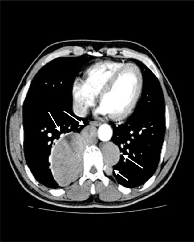
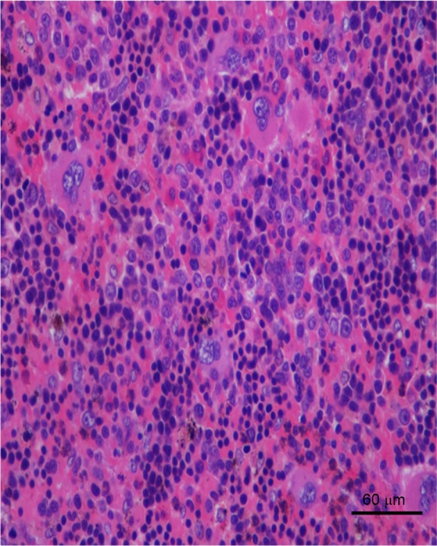
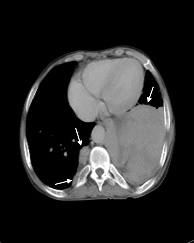
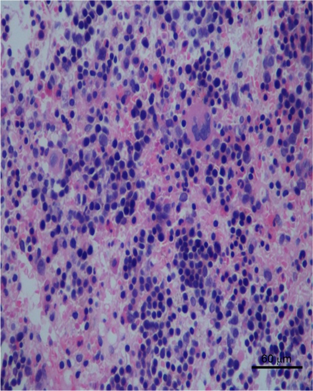
Extramedullary hematopoiesis (EMH) is defined as hematopoiesis occurring in organs outside of the bone marrow. The present report describes two cases of thalassemic patients with paraspinal medullary hematopoiesis and analyzes the clinical manifestations, imaging, pathology, diagnosis and treatment of EMH. In addition, a supplementary review of previously published cases is provided along with a review of the related literature. Computed tomography (CT) of the first case revealed multiple paraspinal masses, and the largest was 6.2×8.0 cm in diameter. Likewise, CT of the second patient revealed multiple paraspinal masses in the bottom of the left thoracic cavity, and the largest was measured 10.1×10.5 cm. The two cases underwent surgical biopsy and the findings were compatible with a diagnosis of EMH. In conclusion, EMH is a compatible and rare disease, and should be distinguished from other neoplasms. EMH must considered when masses with characteristic radiologic appearance are detected in patients with thalassemia intermedia.
Keywords: anemia; bone marrow; extramedullary hematopoisis; neoplasm; thalassemia.
Figures
Similar articles
-
A persistent cough as atypical clinical presentation of intrathoracic extramedullary hematopoiesis (EMH) in a female with thalassemia intermedia.Acta Biomed. 2018 Feb 16;89(2-S):41-46. doi: 10.23750/abm.v89i2-S.7086. Acta Biomed. 2018. PMID: 29451228 Free PMC article.
-
Ectopic extramedullary hematopoiesis: evaluation and treatment of a rare and benign paraspinal/epidural tumor.J Neurosurg Spine. 2013 Mar;18(3):236-42. doi: 10.3171/2012.12.SPINE12720. Epub 2013 Jan 18. J Neurosurg Spine. 2013. PMID: 23330877
-
A Clinical Risk Score for Predicting Paraspinal Extramedullary Hematopoiesis in Patients with Thalassemia: The KKU-EMH Score.J Med Assoc Thai. 2017 Apr;100(4):389-95. J Med Assoc Thai. 2017. PMID: 29911832
-
Paraspinal extramedullary hematopoiesis in hereditary spherocytosis with a concurrent follicular lymphoma: case report and review of the literature.Diagn Pathol. 2015 Sep 15;10:158. doi: 10.1186/s13000-015-0394-x. Diagn Pathol. 2015. PMID: 26369323 Free PMC article. Review.
-
Extramedullary hematopoiesis on 18F-FDG PET/CT in a patient with thalassemia and nasopharyngeal carcinoma: A case report and literature review.J Cancer Res Ther. 2015 Oct-Dec;11(4):1034. doi: 10.4103/0973-1482.150359. J Cancer Res Ther. 2015. PMID: 26881631 Review.
Cited by
-
Thoracic and paraspinal extramedullary hematopoiesis in a cat with chronic non-regenerative anemia.JFMS Open Rep. 2018 Sep 17;4(2):2055116918798868. doi: 10.1177/2055116918798868. eCollection 2018 Jul-Dec. JFMS Open Rep. 2018. PMID: 30245843 Free PMC article.
-
Spinal cord compression secondary to intraspinal extramedullary hematopoiesis.J Family Community Med. 2023 Oct-Dec;30(4):317-319. doi: 10.4103/jfcm.jfcm_151_23. Epub 2023 Oct 13. J Family Community Med. 2023. PMID: 38044968 Free PMC article.
-
[Fine-needle aspiration cell pathology for diagnosis of intrathoracic extramedullary hematopoiesis presenting as a posterior mediastinal tumor: a case report].Nan Fang Yi Ke Da Xue Xue Bao. 2017 May 20;37(5):698-703. doi: 10.3969/j.issn.1673-4254.2017.05.23. Nan Fang Yi Ke Da Xue Xue Bao. 2017. PMID: 28539298 Free PMC article. Chinese.
-
Safety of Repeated Administration of Xenogeneic Human Apoptotic State (Allocetra-OTS) in Sprague Dawley Rats.Pharmaceutics. 2024 Mar 20;16(3):426. doi: 10.3390/pharmaceutics16030426. Pharmaceutics. 2024. PMID: 38543320 Free PMC article.
-
Huge extramedullary hematopoiesis mass in the posterior mediastinum: a case report.Front Oncol. 2024 Dec 13;14:1489785. doi: 10.3389/fonc.2024.1489785. eCollection 2024. Front Oncol. 2024. PMID: 39735597 Free PMC article.
References
-
- Bozzini CE, Rendo ME Barrio, Devoto FC, Epper CE. Studies on medullary and extramedullary erythropoiesis in the adult mouse. Am J Physiol. 1970;219:724–728. - PubMed
-
- Taheri M Sanei, Birang SH, Shahnazi M, Hemadi H. Large splenic mass of extramedullary hematopoiesis. Iran J Radiol. 2005;2:99–101.
LinkOut - more resources
Full Text Sources
Other Literature Sources