

NUCLEI SEGMENTATION VIA SPARSITY CONSTRAINED CONVOLUTIONAL REGRESSION
- PMID: 28101301
- PMCID: PMC5239217
- DOI: 10.1109/ISBI.2015.7164109
NUCLEI SEGMENTATION VIA SPARSITY CONSTRAINED CONVOLUTIONAL REGRESSION
Abstract
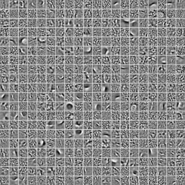
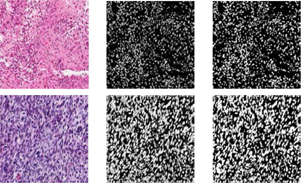
Automated profiling of nuclear architecture, in histology sections, can potentially help predict the clinical outcomes. However, the task is challenging as a result of nuclear pleomorphism and cellular states (e.g., cell fate, cell cycle), which are compounded by the batch effect (e.g., variations in fixation and staining). Present methods, for nuclear segmentation, are based on human-designed features that may not effectively capture intrinsic nuclear architecture. In this paper, we propose a novel approach, called sparsity constrained convolutional regression (SCCR), for nuclei segmentation. Specifically, given raw image patches and the corresponding annotated binary masks, our algorithm jointly learns a bank of convolutional filters and a sparse linear regressor, where the former is used for feature extraction, and the latter aims to produce a likelihood for each pixel being nuclear region or background. During classification, the pixel label is simply determined by a thresholding operation applied on the likelihood map. The method has been evaluated using the benchmark dataset collected from The Cancer Genome Atlas (TCGA). Experimental results demonstrate that our method outperforms traditional nuclei segmentation algorithms and is able to achieve competitive performance compared to the state-of-the-art algorithm built upon human-designed features with biological prior knowledge.
Keywords: H&E tissue section; Nuclear/Background classification; convolutional neural network; sparse coding.
Figures
References
-
- Latson L, Sebek N, Powell K. Automated cell nuclear segmentation in color images of hematoxylin and eosin-stained breast biopsy. Analytical and Quantitative Cytology and Histology. 2003;26(6):321–331. - PubMed
-
- Ballaro B, Florena A, Franco V, Tegolo D, Tripodo C, Valenti C. An automated image analysis methodology for classifying megakaryocytes in chronic myeloproliferative disorders. Medical Image Analysis. 2008;12:703–712. - PubMed
-
- Datar M, Padfield D, Cline H. Color and texture based segmentation of molecular pathology images using HSOMs. ISBI. 2008:292–295.
-
- Fatakdawala H, Xu J, Basavanhally A, Bhanot G, Ganesan S, Feldman F, Tomaszewski J, Madabhushi A. Expectation-maximization-driven geodesic active contours with overlap resolution (EMaGACOR): Application to lymphocyte segmentation on breast cancer histopathology. IEEE Transactions on Biomedical Engineering. 2010;57(7):1676–1690. - PubMed
Grants and funding
LinkOut - more resources
Full Text Sources
Other Literature Sources