. 2017 Jan 13:5:1.
doi: 10.1186/s40170-016-0163-7. eCollection 2017.

Adipocyte lipolysis links obesity to breast cancer growth: adipocyte-derived fatty acids drive breast cancer cell proliferation and migration

Affiliations

Adipocyte lipolysis links obesity to breast cancer growth: adipocyte-derived fatty acids drive breast cancer cell proliferation and migration

Seher Balaban et al. Cancer Metab. .

Abstract

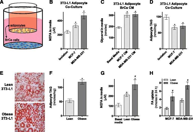
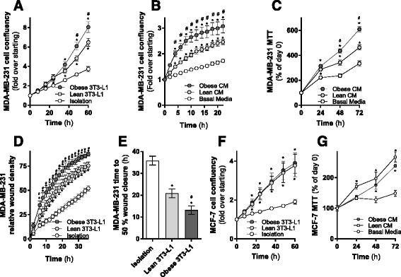
Background: Obesity is associated with increased recurrence and reduced survival of breast cancer. Adipocytes constitute a significant component of breast tissue, yet their role in provisioning metabolic substrates to support breast cancer progression is poorly understood.

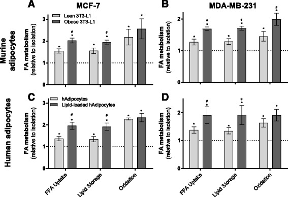
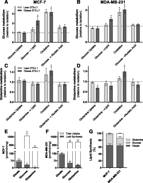
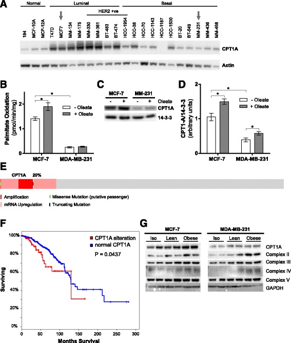
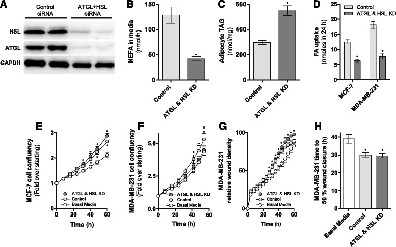
Results: Here, we show that co-culture of breast cancer cells with adipocytes revealed cancer cell-stimulated depletion of adipocyte triacylglycerol. Adipocyte-derived free fatty acids were transferred to breast cancer cells, driving fatty acid metabolism via increased CPT1A and electron transport chain complex protein levels, resulting in increased proliferation and migration. Notably, fatty acid transfer to breast cancer cells was enhanced from "obese" adipocytes, concomitant with increased stimulation of cancer cell proliferation and migration. This adipocyte-stimulated breast cancer cell proliferation was dependent on lipolytic processes since HSL/ATGL knockdown attenuated cancer cell responses.

Conclusions: These findings highlight a novel and potentially important role for adipocyte lipolysis in the provision of metabolic substrates to breast cancer cells, thereby supporting cancer progression.

Keywords: Adipocytes; Breast cancer; Lipid metabolism; Metabolic crosstalk; Obesity.


Figures

Fig. 1
Fig. 1
Breast cancer cells stimulate lipolysis in mature 3T3-L1 adipocytes. a Schematic of co-culture approach. b Media non-esterified fatty acids (NEFA) concentration following co-culture of 3T3-L1 adipocytes without (isolation) or with MCF-7 and MDA-MB-231 cells for 24 h (four independent experiments performed in triplicate). c Glycerol release from 3T3-L1 adipocytes incubated in basal media, MCF-7-conditioned media (CM), and MDA-MB-231-conditioned media at the end of 24 h incubation (seven independent experiments performed in quadruplicate). d 3T3-L1 adipocytes triacylglycerol (TAG) content after 48 h co-culture with or without MCF-7 or MDA-MB-231 cells (two independent experiments performed in triplicate). e 3T3-L1 adipocyte Oil Red O staining of lipid droplets, f TAG content (three independent experiments performed in triplicate), and g NEFA release from lean (normal media) and obese (1 mM fatty acid mixture for 24 h) adipocytes compared to basal media levels (four independent experiments performed in duplicate). h Transfer of adipocyte-derived 3H-fatty acids from lean or obese 3T3-L1 adipocytes to MCF-7 and MDA-MB-231 cells (three independent experiments performed in duplicate). Data are presented as mean ± SEM. *P ≤ 0.05 vs. controls; #P ≤ 0.05 vs. lean. bd and g By one-way ANOVA followed by Tukey’s multiple comparisons test, f, h by Student’s t test
Fig. 2
Fig. 2
Adipocytes alter fatty acid partitioning in breast cancer cells. a MCF-7 cells and b MDA-MB-231 cells [1-14C]-oleate metabolism including total uptake (sum of media 14CO2, 14C activity in both the aqueous and organic phases of a Folch extraction), incorporation into intracellular lipids (storage), and 14CO2 generation (oxidation) after co-culture with or without 3T3-L1 adipocytes for 48 h (three independent experiments performed in triplicate). c MCF-7 cells and d MDA-MB-231 cells [1-14C]-oleate metabolism after co-culture with or without differentiated human primary mammary pre-adipocytes for 48 h (two independent experiments performed in duplicate). Data are presented as mean ± SEM, relative to cells in isolation (dotted line). *P ≤ 0.05 compared to isolation; #P ≤ 0.05 compared to lean by one-way ANOVA followed by Tukey’s multiple comparisons test
Fig. 3
Fig. 3
Altered glucose and glutamine metabolism in MDA-MB-231 and MCF-7 cells following co-culture with lean and obese adipocytes a MCF-7 cells and b MDA-MB-231 cells [U-14C]-glucose metabolism including total uptake (sum of media 14CO2, 14C activity in both the aqueous and organic phases of a Folch extraction), incorporation into intracellular lipids, 14CO2 generation (oxidation), and incorporation into DNA/RNA after co-cultured with or without 3T3-L1 adipocytes for 48 h (three independent experiments performed in duplicate, relative to cells in isolation (dotted line)). c MCF-7 cells and d MDA-MB-231 cells [1-14C]-l-glutamine metabolism after co-cultured with or without 3T3-L1 adipocytes for 48 h (three independent experiments performed in duplicate, relative to cells in isolation (dotted line)). e, f Absolute rates of 14C-labeled substrate incorporation into intracellular lipids (lipid synthesis) and total uptake (sum of media 14CO2, 14C activity in both the aqueous and organic phases of a Folsch extraction) of various substrates in MCF-7 (e) and MDA-MB-231 (f) cells and g percent contribution of substrates to lipid synthesis in both cell lines in the basal state (three independent experiments performed in duplicate). Data are presented as mean ± SEM. ad *P ≤ 0.05 compared to isolation; #P ≤ 0.05 compared to lean by one-way ANOVA followed by Tukey’s multiple comparisons test. ef *P ≤ 0.05 compared to oleate; #P ≤ 0.05 compared to glucose by one-way ANOVA followed by Tukey’s multiple comparisons test
Fig. 4
Fig. 4
MCF-7 cells have greater CPT1 protein levels and fatty acid oxidation rates compared to MDA-MB-231 cells, and these are increased high lipid environments. a CPT1A expression in a range of normal, luminal, and basal breast cancer cell lines. b [1-14C]-palmitate oxidation, c representative immunoblots of three independent experiments, and d densitometric quantitation of CPT1A protein levels in MCF-7 and MDA-MB-231 cells in the basal state and after 24 h culture in 0.3 mM oleate. e Oncoprint output showing frequency of CPT1A alteration in a TCGA cohort of ER+ breast cancer (n = 594) (red rectangle, amplification; light red rectangle, mRNA upregulation; black rectangle, truncated mutation; green rectangle, missense mutation; gray rectangle, unaltered). f Overall survival among ER+ breast carcinoma TCGA cases based on CPT1A alterations. g Representative immunoblots of CPT1A and protein subunits in the mitochondrial complexes (complex II-30 kDa, complex III-Core protein 2, complex IV-subunit 1, and complex V-alpha subunit) in MCF-7 and MDA-MB-231 cells co-cultured with or without lean or obese 3T3-L1 adipocytes (three independent experiments performed in duplicate). Data are presented as mean ± SEM. *P ≤ 0.05 by two-way ANOVA followed by Tukey’s multiple comparisons test
Fig. 5
Fig. 5
Adipocytes enhance breast cancer cells proliferation and migration rate. a MDA-MB-231GFP cell proliferation co-cultured with or without lean or obese 3T3-L1 adipocytes (three independent experiments performed in quadruplicate). b IncuCyte and c MTT assessment of MDA-MB-231 cell proliferation cultured in conditioned media from lean or obese 3T3-L1 adipocytes (three independent experiments performed in quadruplicate). d, e Migration of MDA-MB-231 cells co-cultured with or without lean or obese 3T3-L1 adipocytes (three independent experiments performed in quadruplicate). f MCF-7GFP cell proliferation co-cultured with or without lean or obese 3T3-L1 adipocytes (three independent experiments performed in quadruplicate). g MTT assessment of MCF-7 cell proliferation cultured in conditioned media from lean or obese 3T3-L1 adipocytes (three independent experiments performed in quadruplicate). Data are presented as mean ± SEM. *P ≤ 0.05 vs. controls; #P ≤ 0.05 vs. lean ad and f, g by two-way ANOVA repeated measure followed by Tukey’s multiple comparisons test and e by one-way ANOVA followed by Tukey’s multiple comparisons test
Fig. 6
Fig. 6
siRNA-mediated knockdown of ATGL and HSL in 3T3-L1 adipocytes. a Representative immunoblots of ATGL and HSL knockdown in 3T3-L1 adipocytes. Image is representative of six independent experiments. b NEFA secretion and c TAG content in 3T3-L1 adipocytes electroporated with either non-targeting (control) or LIPE (HSL) and PNPLA2 (ATGL) siRNAs (four independent experiments performed in triplicate). d Transfer of adipocyte-derived 3H-fatty acids to MCF-7 and MDA-MB-231 cells (three independent experiments performed in duplicate). e MDA-MB-231 and f MCF-7 cell proliferation co-cultured with or without ATGL/HSL knockdown 3T3-L1 adipocytes (three independent experiments performed in quadruplicate). g, h Migration of MDA-MB-231 cells co-cultured with or without ATGL/HSL knockdown 3T3-L1 adipocytes (three independent experiments performed in quadruplicate). Data are means and SEM. bd *P < 0.05 compared to control siRNA with Student’s t test. eg *P ≤ 0.05 vs. basal media, #P ≤ 0.05 compared to ATGL and HSL KD by two-way ANOVA repeated measures followed by Tukey’s multiple comparisons test and h *P ≤ 0.05 vs. basal media by one-way ANOVA followed by Tukey’s multiple comparisons test

References

    1. Martinez-Outschoorn UE, Peiris-Pages M, Pestell RG, Sotgia F, Lisanti MP. Cancer metabolism: a therapeutic perspective. Nat Rev Clin Oncol. 2017;14(1):11-31. doi:10.1038/nrclinonc.2016.60. [PMID: 27141887]. - PubMed
    1. Martinez-Outschoorn U, Sotgia F, Lisanti MP. Tumor microenvironment and metabolic synergy in breast cancers: critical importance of mitochondrial fuels and function. Semin Oncol. 2014;41:195–216. doi: 10.1053/j.seminoncol.2014.03.002. - DOI - PubMed
    1. Zhang DH, Tai LK, Wong LL, Chiu LL, Sethi SK, Koay ESC. Proteomic study reveals that proteins involved in metabolic and detoxification pathways are highly expressed in HER-2/neu-positive breast cancer. Mol Cell Proteomics. 2005;4:1686–96. doi: 10.1074/mcp.M400221-MCP200. - DOI - PubMed
    1. Yoon S, Lee MY, Park SW, Moon JS, Koh YK, Ahn YH, Park BW, Kim KS. Up-regulation of Acetyl-CoA carboxylase alpha and fatty acid synthase by human epidermal growth factor receptor 2 at the translational level in breast cancer cells. J Biol Chem. 2007;282:26122–31. doi: 10.1074/jbc.M702854200. - DOI - PubMed
    1. Zaidi N, Lupien L, Kuemmerle NB, Kinlaw WB, Swinnen JV, Smans K. Lipogenesis and lipolysis: the pathways exploited by the cancer cells to acquire fatty acids. Prog Lipid Res. 2013;52:585–9. doi: 10.1016/j.plipres.2013.08.005. - DOI - PMC - PubMed