

Detection and classification of three-class initial dips from prefrontal cortex
- PMID: 28101424
- PMCID: PMC5231305
- DOI: 10.1364/BOE.8.000367
Detection and classification of three-class initial dips from prefrontal cortex
Abstract
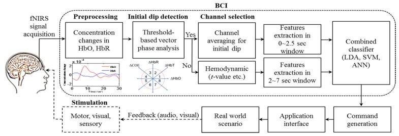
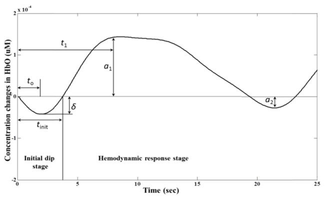
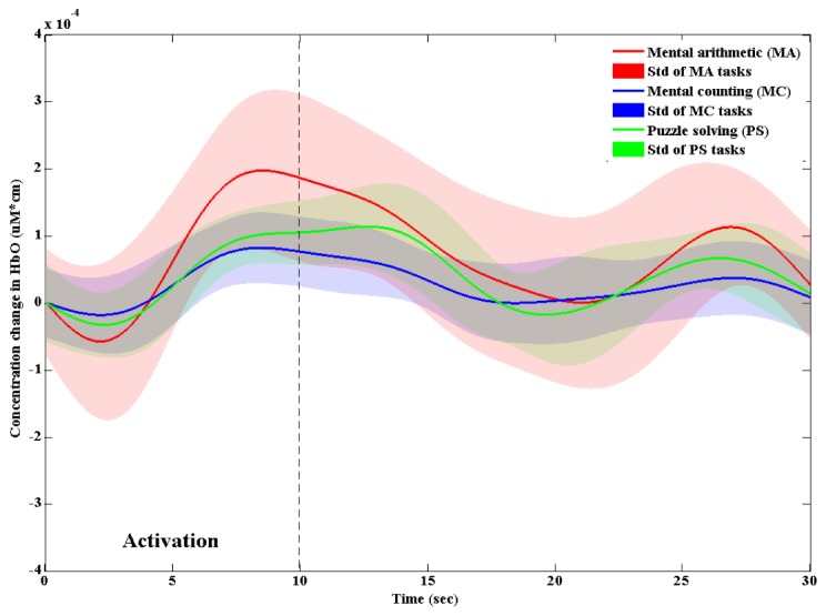
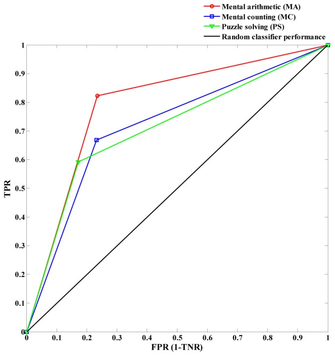
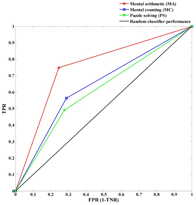
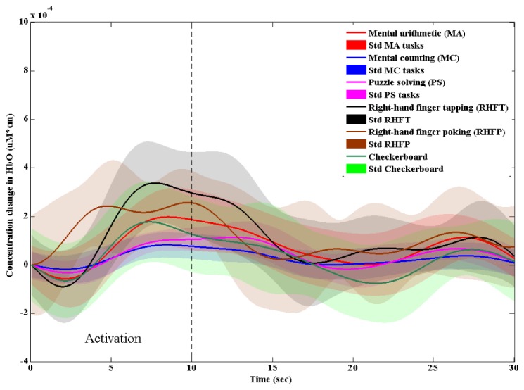
In this paper, the use of initial dips using functional near-infrared spectroscopy (fNIRS) for brain-computer interface (BCI) is investigated. Features and window sizes for detecting initial dips are also discussed. Three mental tasks including mental arithmetic, mental counting, and puzzle solving are performed in obtaining fNIRS signals from the prefrontal cortex. Vector-based phase analysis method combined with a threshold circle, as a decision criterion, are used to detect the initial dips. Eight healthy subjects participate in experiment. Linear discriminant analysis is used as a classifier. To classify initial dips, five features (signal mean, peak value, signal slope, skewness, and kurtosis) of oxy-hemoglobin (HbO) and four different window sizes (0~1, 0~1.5, 0~2, and 0~2.5 sec) are examined. It is shown that a combination of signal mean and peak value and a time period of 0~2.5 sec provide the best average classification accuracy of 57.5% for three classes. To further validate the result, three-class classification using the conventional hemodynamic response (HR) is also performed, in which two features (signal mean and signal slope) and 2~7 sec window size have yielded the average classification accuracy of 65.9%. This reveals that fNIRS-based BCI using initial dip detection can reduce the command generation time from 7 sec to 2.5 sec while the classification accuracy is a bit sacrificed from 65.9% to 57.5% for three mental tasks. Further improvement can be made by using deoxy hemoglobin signals in coping with the slow HR problem.
Keywords: (070.5010) Pattern recognition; (170.2655) Functional monitoring and imaging; (200.3050) Information processing; (300.0300) Spectroscopy.
Figures
Similar articles
-
Neuronal Activation Detection Using Vector Phase Analysis with Dual Threshold Circles: A Functional Near-Infrared Spectroscopy Study.Int J Neural Syst. 2018 Dec;28(10):1850031. doi: 10.1142/S0129065718500314. Epub 2018 Jun 24. Int J Neural Syst. 2018. PMID: 30045647
-
Determining Optimal Feature-Combination for LDA Classification of Functional Near-Infrared Spectroscopy Signals in Brain-Computer Interface Application.Front Hum Neurosci. 2016 May 25;10:237. doi: 10.3389/fnhum.2016.00237. eCollection 2016. Front Hum Neurosci. 2016. PMID: 27252637 Free PMC article.
-
Reduction of Delay in Detecting Initial Dips from Functional Near-Infrared Spectroscopy Signals Using Vector-Based Phase Analysis.Int J Neural Syst. 2016 May;26(3):1650012. doi: 10.1142/S012906571650012X. Epub 2016 Jan 14. Int J Neural Syst. 2016. PMID: 26971785
-
fNIRS-based brain-computer interfaces: a review.Front Hum Neurosci. 2015 Jan 28;9:3. doi: 10.3389/fnhum.2015.00003. eCollection 2015. Front Hum Neurosci. 2015. PMID: 25674060 Free PMC article. Review.
-
Feature Extraction and Classification Methods for Hybrid fNIRS-EEG Brain-Computer Interfaces.Front Hum Neurosci. 2018 Jun 28;12:246. doi: 10.3389/fnhum.2018.00246. eCollection 2018. Front Hum Neurosci. 2018. PMID: 30002623 Free PMC article. Review.
Cited by
-
Early Detection of Alzheimer's Disease Using Non-invasive Near-Infrared Spectroscopy.Front Aging Neurosci. 2018 Nov 9;10:366. doi: 10.3389/fnagi.2018.00366. eCollection 2018. Front Aging Neurosci. 2018. PMID: 30473662 Free PMC article.
-
Effects of Acupuncture Therapy on MCI Patients Using Functional Near-Infrared Spectroscopy.Front Aging Neurosci. 2019 Aug 30;11:237. doi: 10.3389/fnagi.2019.00237. eCollection 2019. Front Aging Neurosci. 2019. PMID: 31543811 Free PMC article.
-
Brain-computer interface to predict impulse buying behavior using functional near-infrared spectroscopy.Sci Rep. 2022 Oct 26;12(1):18024. doi: 10.1038/s41598-022-22653-8. Sci Rep. 2022. PMID: 36289356 Free PMC article.
-
An fNIRS-based investigation of visual merchandising displays for fashion stores.PLoS One. 2018 Dec 11;13(12):e0208843. doi: 10.1371/journal.pone.0208843. eCollection 2018. PLoS One. 2018. PMID: 30533055 Free PMC article.
-
Detection of Mild Cognitive Impairment Using Convolutional Neural Network: Temporal-Feature Maps of Functional Near-Infrared Spectroscopy.Front Aging Neurosci. 2020 May 21;12:141. doi: 10.3389/fnagi.2020.00141. eCollection 2020. Front Aging Neurosci. 2020. PMID: 32508627 Free PMC article.
References
-
- Frostig R. D., Lieke E. E., Ts’o D. Y., Grinvald A., “Cortical functional architecture and local coupling between neuronal activity and the microcirculation revealed by in vivo high-resolution optical imaging of intrinsic signals,” Proc. Natl. Acad. Sci. U.S.A. 87(16), 6082–6086 (1990).10.1073/pnas.87.16.6082 - DOI - PMC - PubMed
-
- Menon R. S., Ogawa S., Hu X., Strupp J. P., Anderson P., Uğurbil K., “BOLD based functional MRI at 4 Tesla includes a capillary bed contribution: echo-planar imaging correlates with previous optical imaging using intrinsic signals,” Magn. Reson. Med. 33(3), 453–459 (1995).10.1002/mrm.1910330323 - DOI - PubMed
LinkOut - more resources
Full Text Sources
Other Literature Sources