. 2016 Dec 23;8(1):460-474.
doi: 10.1364/BOE.8.000460. eCollection 2017 Jan 1.

Non-invasive terahertz imaging of tissue water content for flap viability assessment

Affiliations

Non-invasive terahertz imaging of tissue water content for flap viability assessment

Neha Bajwa et al. Biomed Opt Express. .

Abstract

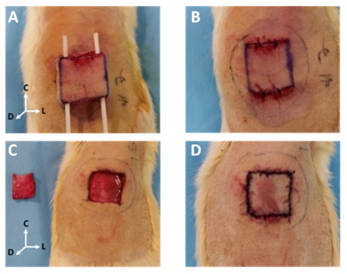
Accurate and early prediction of tissue viability is the most significant determinant of tissue flap survival in reconstructive surgery. Perturbation in tissue water content (TWC) is a generic component of the tissue response to such surgeries, and, therefore, may be an important diagnostic target for assessing the extent of flap viability in vivo. We have previously shown that reflective terahertz (THz) imaging, a non-ionizing technique, can generate spatially resolved maps of TWC in superficial soft tissues, such as cornea and wounds, on the order of minutes. Herein, we report the first in vivo pilot study to investigate the utility of reflective THz TWC imaging for early assessment of skin flap viability. We obtained longitudinal visible and reflective THz imagery comparing 3 bipedicled flaps (i.e. survival model) and 3 fully excised flaps (i.e. failure model) in the dorsal skin of rats over a postoperative period of 7 days. While visual differences between both models manifested 48 hr after surgery, statistically significant (p < 0.05, independent t-test) local differences in TWC contrast were evident in THz flap image sets as early as 24 hr. Excised flaps, histologically confirmed as necrotic, demonstrated a significant, yet localized, reduction in TWC in the flap region compared to non-traumatized skin. In contrast, bipedicled flaps, histologically verified as viable, displayed mostly uniform, unperturbed TWC across the flap tissue. These results indicate the practical potential of THz TWC sensing to accurately predict flap failure 24 hours earlier than clinical examination.

Keywords: (110.0110) Imaging systems; (170.0170) Medical optics and biotechnology; (170.6795) Terahertz imaging.


Figures

Fig. 1
Fig. 1
In vivo THz imaging system. (a) Visible image of the reflective THz system. Dorsal flaps of anesthetized rats are raster scanned on x- and y-stepper motors beneath the imager. The encirlcled optics correspond to (b) off-axis parabolic (OAP) mirrors used to focus the THz beam onto a thin-film Mylar window that serves to mitigate surface contours of the underlying flap.
Fig. 2
Fig. 2
Myocutaneous flap design. (a) An axially-based bipedicled flap (i.e. survival model), measuring 2.5 x 2.5 cm2 in area with cephalic and caudal incisions, that is elevated deep to the panniculus carnosous and then (b) surgically secured in its anatomical position. (c) Completely excised myocutaneous skin flap (i.e. failure model), measuring 2.5 x 2.5 cm2 in area with circumferential flap excision, that is elevated deep to the panniculus carnosous and (d) sutured back in anatomic position. “L,” “D,” and “C” correspond to lateral, dorsal, and cephalic anatomical directions of the rat.
Fig. 3
Fig. 3
Histology design. Histological evaluation of tissue viability for (a) an excised flap and (b) a bipedicled flap in a rat imaged under a Mylar window (denoted by the solid black circle). A zoom-in diagram shows the field of view (FOV) of the window, which captures the flap tissue (hatched red region), incisions (solid red lines), and non-traumatized tissue (surrounding tan region. Sagittal histological slices of 5 µm thickness were taken from 2 cm x 2 mm tissue sections (dotted green rectangle) harvested from the cephalic incision site (solid red arrow) on day 7. Dotted red arrow denotes the lateral incision. A suture in each slice (black loop) served as an intradermal fiducial marker for image registration of histology to the same region in the FOV captured by THz-time series imagery. “L,” “D,” and “C” correspond to anatomical directions detailed in Fig. 2.
Fig. 4
Fig. 4
In vivo visible and THz time-series flap imagery. (a) Excised flaps and (b) bipedicled flaps in the dorsum of anesthetized rats were imaged under a 12.7 µm Mylar window over a 7-day period. Uninjured tissue and flap tissue are labeled in yellow and gray fields, respectively, in visible images acquired at 15min following surgery. The hot color map of THz imagery transforms black to the global minimum THz reflectivity and white to the global maximum THz reflectivity. “L,” “D,” and “C” in the 3D axis denote directions detailed in Fig. 2. Black scale bars represent ~1 cm in the FOV. ~1.5 cm long contours, indicated by solid white horizontal lines, segment the cephalic incision, flap region, and non-traumatized tissue. Solid white arrows locate the leakage of blood, indicated by high THz reflectivity, from the incision site.
Fig. 5
Fig. 5
Spatial THz reflectivity profiles for THz flap image sets. (a) Profiles of %∆ THz reflectivity in excised flaps and (b) bipedicled flaps with respect to pre-surgery values were generated for white contours that segment THz images in Fig. 4. These THz profiles captured the cephalic incision (i.e. dashed vertical lines), flap region (i.e. left of incision), and non-traumatized tissue (i.e. right of incision) of tissue flaps at 15min, 24hr, and 48hr post-operation. All red arrows denote increased THz reflectivity due to the presence of blood immediately following surgery, a confounder to THz contrast. (c) Pixel-by-pixel differences in THz reflectivity between both flap models at 24hr following surgery were confirmed to be statistically significant (p <0.05, the solid red line) across the entire length of the white contour (i.e. x-axis) using an independent student t-test.
Fig. 6
Fig. 6
Clinical and histological assessment of flap viability. Post-operative visual surveillance of (a) excised flaps and (b) bipedicled flaps over 7 days. “L,” “D,” and “C” correspond to directions in Fig. 2. (c) 1X and 2X representative hematoxylin and eosin (H&E) staining for tissue flap margins spatially map to the cephalic incision at day 7, demarcated by white horizontal lines in the visible imagery. In the H&E image, flap tissue and non-traumatized tissue lie to the left and right of the incision site (denoted by a black arrow), respectively. Necrotic tissue appears dark magenta in the H&E stain. The black scale bar and red scale bar represent ~1 mm and ~450 µm, respectively.

References

    1. Payette J. R., Kohlenberg E., Leonardi L., Pabbies A., Kerr P., Liu K.-Z., Sowa M. G., “Assessment of Skin Flaps Using Optically Based Methods for Measuring Blood Flow and Oxygenation,” Plast. Reconstr. Surg. 115(2), 539–546 (2005).10.1097/01.PRS.0000148415.54546.CA - DOI - PubMed
    1. Bergkvist M., Henricson J., Iredahl F., Tesselaar E., Sjöberg F., Farnebo S., “Assessment of microcirculation of the skin using Tissue Viability Imaging: A promising technique for detecting venous stasis in the skin,” Microvasc. Res. 101, 20–25 (2015).10.1016/j.mvr.2015.06.002 - DOI - PubMed
    1. Di Sieno L., Bettega G., Berger M., Hamou C., Aribert M., Mora A. D., Puszka A., Grateau H., Contini D., Hervé L., Coll J.-L., Dinten J.-M., Pifferi A., Planat-Chrétien A., “Toward noninvasive assessment of flap viability with time-resolved diffuse optical tomography: a preclinical test on rats,” J. Biomed. Opt. 21(2), 025004 (2016).10.1117/1.JBO.21.2.025004 - DOI - PubMed
    1. Yang Q., Ren Z. H., Chickooree D., Wu H. J., Tan H. Y., Wang K., He Z. J., Gong C. J., Ram V., Zhang S., “The effect of early detection of anterolateral thigh free flap crisis on the salvage success rate, based on 10 years of experience and 1072 flaps,” Int. J. Oral Maxillofac. Surg. 43(9), 1059–1063 (2014).10.1016/j.ijom.2014.06.003 - DOI - PubMed
    1. Chen K.-T., Mardini S., Chuang D. C.-C., Lin C.-H., Cheng M.-H., Lin Y.-T., Huang W.-C., Tsao C.-K., Wei F.-C., “Timing of presentation of the first signs of vascular compromise dictates the salvage outcome of free flap transfers,” Plast. Reconstr. Surg. 120(1), 187–195 (2007).10.1097/01.prs.0000264077.07779.50 - DOI - PubMed