. 2017 Jan 11;2(1):e00375-16.
doi: 10.1128/mSphere.00375-16. eCollection 2017 Jan-Feb.

Host Range Restriction of Insect-Specific Flaviviruses Occurs at Several Levels of the Viral Life Cycle

Affiliations

Host Range Restriction of Insect-Specific Flaviviruses Occurs at Several Levels of the Viral Life Cycle

Sandra Junglen et al. mSphere. .

Abstract

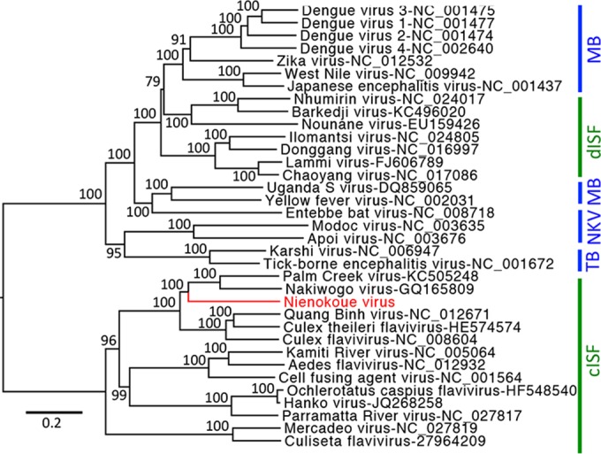
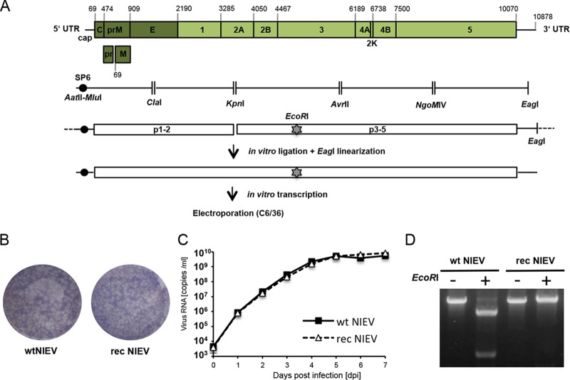
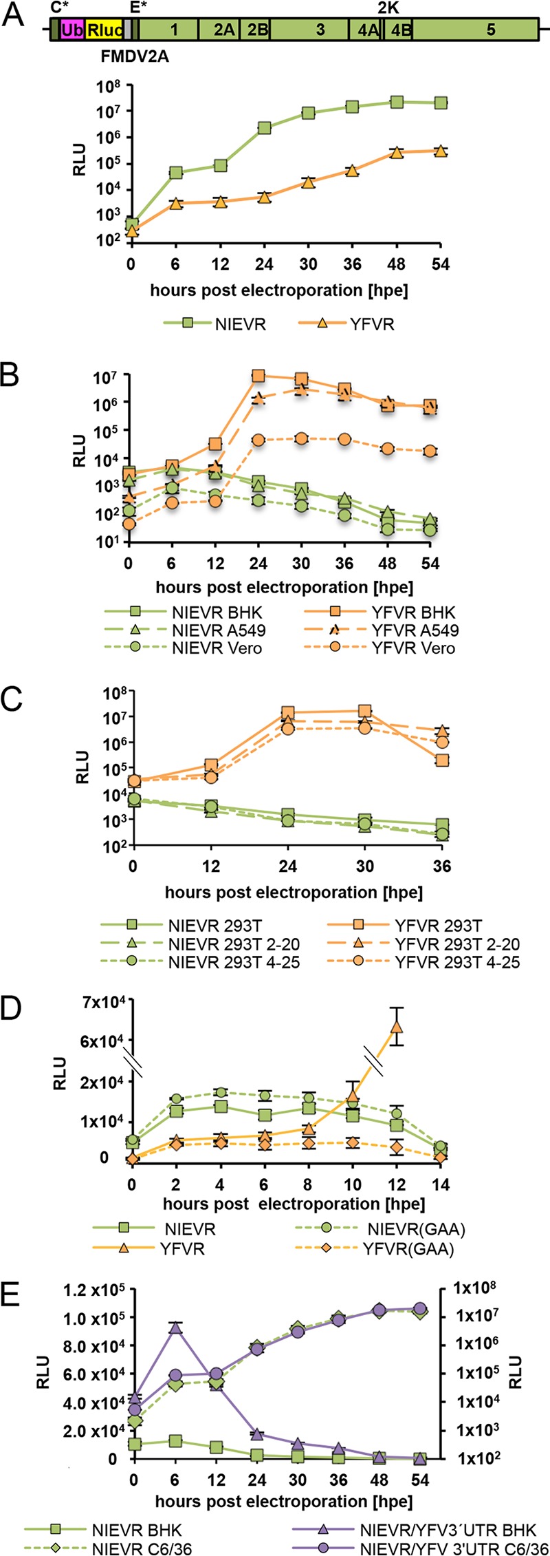
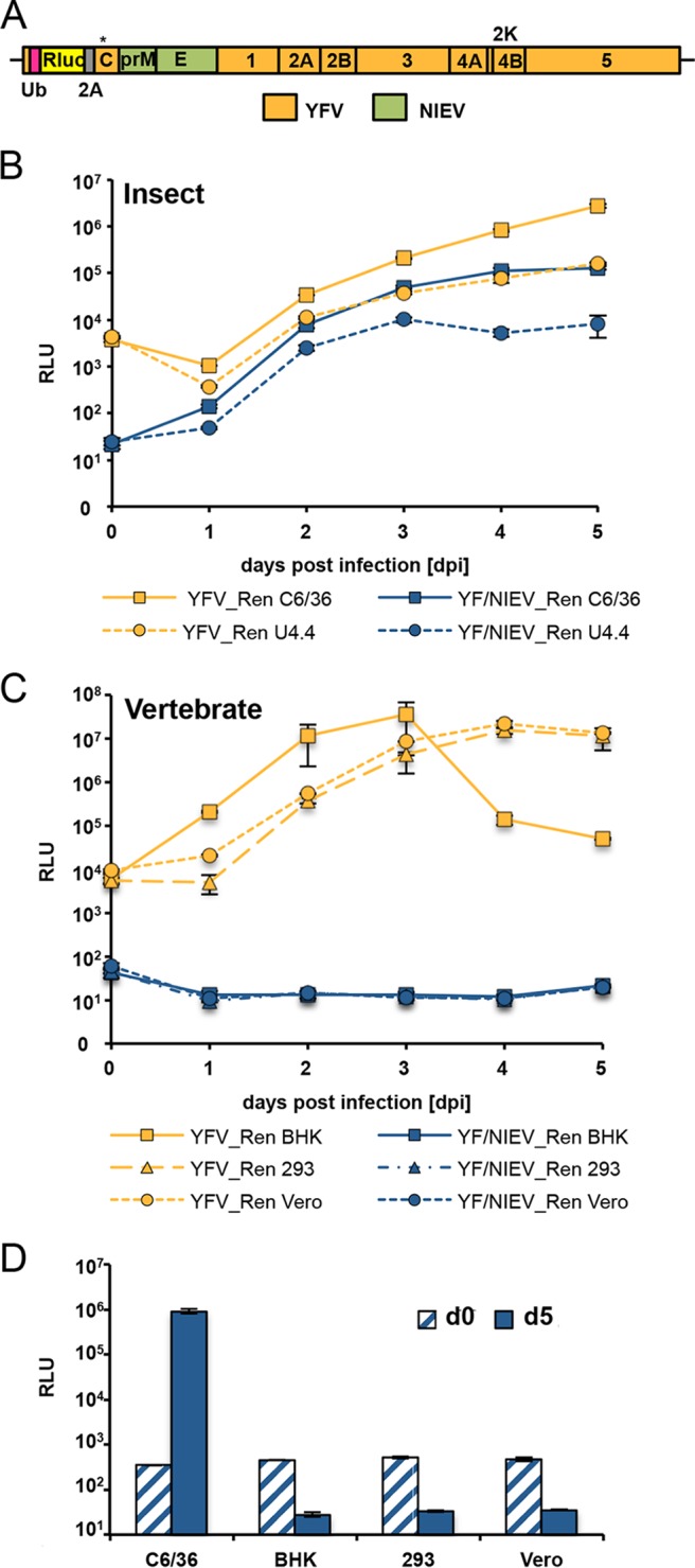
The genus Flavivirus contains emerging arthropod-borne viruses (arboviruses) infecting vertebrates, as well as insect-specific viruses (ISVs) (i.e., viruses whose host range is restricted to insects). ISVs are evolutionary precursors to arboviruses. Knowledge of the nature of the ISV infection block in vertebrates could identify functions necessary for the expansion of the host range toward vertebrates. Mapping of host restrictions by complementation of ISV and arbovirus genome functions could generate knowledge critical to predicting arbovirus emergence. Here we isolated a novel flavivirus, termed Niénokoué virus (NIEV), from mosquitoes sampled in Côte d'Ivoire. NIEV groups with insect-specific flaviviruses (ISFs) in phylogeny and grows in insect cells but not in vertebrate cells. We generated an infectious NIEV cDNA clone and a NIEV reporter replicon to study growth restrictions of NIEV in comparison to yellow fever virus (YFV), for which the same tools are available. Efficient RNA replication of the NIEV reporter replicon was observed in insect cells but not in vertebrate cells. Initial translation of the input replicon RNA in vertebrate cells was functional, but RNA replication did not occur. Chimeric YFV carrying the envelope proteins of NIEV was recovered via electroporation in C6/36 insect cells but did not infect vertebrate cells, indicating a block at the level of entry. Since the YF/NIEV chimera readily produced infectious particles in insect cells but not in vertebrate cells despite efficient RNA replication, restriction is also determined at the level of assembly/release. Taking the results together, the ability of ISF to infect vertebrates is blocked at several levels, including attachment/entry and RNA replication as well as assembly/release. IMPORTANCE Most viruses of the genus Flavivirus, e.g., YFV and dengue virus, are mosquito borne and transmitted to vertebrates during blood feeding of mosquitoes. Within the last decade, an increasing number of viruses with a host range exclusively restricted to insects in close relationship to the vertebrate-pathogenic flaviviruses were discovered in mosquitoes. To identify barriers that could block the arboviral vertebrate tropism, we set out to identify the steps at which the ISF replication cycle fails in vertebrates. Our studies revealed blocks at several levels, suggesting that flavivirus host range expansion from insects to vertebrates was a complex process that involved overcoming multiple barriers.

Keywords: host range restriction; infection barriers; insect-specific flavivirus.


Figures

FIG 1
FIG 1
Phylogenetic relationships of NIEV. Complete flavivirus polyproteins were aligned in geneious using MAFFT and the E-INS-I algorithm. Alignment columns were stripped to contain gaps of less than 20%. Maximum likelihood analyses were inferred with the LG substitution matrix and 1,000 bootstrap replicates using PhyML. Nodes are labeled with bootstrap support using percentage values. Vertebrate-pathogenic viruses are labeled with blue vertical lines and insect-specific viruses with green. Abbreviations are as follows: MB, mosquito-borne flaviviruses; TB, tick-borne flaviviruses; NKV, flaviviruses with no known vector; dISF, dual-host-affiliated insect-specific flaviviruses; cISF, classical insect-specific flaviviruses.
FIG 2
FIG 2
Generation and in vitro characterization of recombinant NIEV. (A) Schematic drawing of the NIEV genome organization and reverse genetics system. Depicted nucleotide positions indicate the borders of the encoded viral proteins or the genome end. Below the NIEV genome organization, the reverse-transcribed NIEV cDNA fragments, including the restriction sites used for cloning, are shown. An SP6 promoter sequence was fused to the 5′ end of the NIEV genome. EcoRI indicates the silent genetic marker mutation resulting in deletion of an EcoRI restriction site. After assembly of the cDNA fragments into two plasmids, in vitro ligation was performed followed by EagI linearization of the ligation product. The latter was transcribed in vitro, and the transcribed RNA was electroporated into C6/36 cells to recover rec NIEV. (B) Plaque morphology of wt NIEV and rec NIEV. Plaque morphology was analyzed by ICA in C6/36 cells using a tragacanth overlay. At 7 days postelectroporation, cells were fixed and subjected to crystal violet staining. (C) Growth kinetics of wt NIEV and rec NIEV. C6/36 cells were infected at an MOI of 0.1. Quantification of viral genome copies in the supernatant was performed by real-time PCR. Data represent means and ranges of results of duplicate infection experiments. (D) Verification of the genetic marker introduced into rec NIEV. Viral RNA was isolated from supernatants of cells infected with the indicated viruses and used as the template for RT-PCR spanning the region containing the deleted EcoRI site in rec NIEV. RT-PCR products were loaded either directly (−) or after EcoRI restriction (+) on an ethidium bromide-stained agarose gel.
FIG 3
FIG 3
Analyses of NIEV replication and translation. (A) Top: schematic drawing of the NIEV Renilla replicon. The narrow dark gray boxes represent the remaining structural proteins, namely, the first 30 amino acids of the capsid protein (C*) and the last 23 amino acids of the E protein (E*), which serve as signal sequence for the following NS1 protein. The pink box indicates the ubiquitin protein (Ub) responsible for generation of the N terminus of the Renilla luciferase protein (Rluc; yellow box). The narrow gray box represents the 17-amino-acid-residue autoproteolytic peptide from the foot-and-mouth disease virus (FMDV2A), which cleaves at its own C terminus. Lines indicate the 5′ and 3′ UTRs. Bottom: replication of the NIEV Renilla replicon (NIEVR) in insect cells. C6/36 cells were electroporated with NIEVR RNA transcribed in vitro. For comparison, electroporation of YFV Renilla replicon RNA (YFVR) was performed. Cells were incubated at 28°C, and replication kinetics were monitored by analysis of replicon-derived Renilla luciferase expression at the indicated times p.e. Data represent means and ranges of results of duplicate electroporation experiments presented in this panel and in panels B to D. (B) Replication of NIEVR and YFVR in vertebrate cells. The indicated different vertebrate cells were electroporated with in vitro-transcribed NIEVR and YFVR RNAs. Cells were incubated at 37°C, and replication kinetics were monitored as described for panel A. (C) Replication of NIEVR and YFVR in Dicer- and miRNA-deficient 293T cells. Wild-type 293T cells or Dicer- and miRNA-deficient cell clones 2–20 or 4–25 were electroporated with in vitro-transcribed NIEVR and YFVR RNAs. Cells were incubated at 37°C, and replication kinetics were monitored as described for panel A. (D) Analysis of NIEVR translation. BHK cells were electroporated with replication-incompetent Renilla replicon RNAs bearing the exchange of the polymerase GDD motif to GAA, resulting in NIEVR(GAA) or YFVR(GAA). Wild-type NIEVR and YFVR RNAs were electroporated for comparison. Cells were incubated at 37°C, and replication kinetics were monitored as described for panel A. (E) Analysis of NIEVR/YFV 3′ UTR translation. BHK or C6/36 cells were electroporated with NIEVR containing a YFV 3′ UTR insertion downstream of its ORF stop codon. Wild-type NIEVR was electroporated for comparison. Replication kinetics were monitored as described for panel A. Data represent means and ranges of results of duplicate electroporation experiments. Left axis, BHK; right axis, C6/36.
FIG 4
FIG 4
Construction and characterization of YF/NIEV chimera. (A) Schematic drawing of the YF/NIEV chimera. The envelope proteins prM and E of YFV were exchanged against the corresponding prM and E proteins of NIEV (green boxes). (B) Plaque morphology of YFV and YF/NIEV. C6/36 cells were electroporated with in vitro RNA transcripts of the indicated constructs or were mock transfected and processed for infectious center assay analyses. Seeded cells were overlaid with tragacanth. At 7 days p.e., cells were fixed and subjected to crystal violet staining. (C) Growth kinetics of YFV and YF/NIEV. C6/36 cells were infected at an MOI of 0.1. Quantification of viral genome copies in the supernatant was performed by real-time PCR. Data represent means and ranges of results of duplicate infection experiments. (D) Immunofluorescence analyses after infection of different cells. C6/36 or BHK cells were infected with the indicated viruses at an MOI of 0.1. At 48 h p.i., immunofluorescence analysis was performed using a monoclonal anti-YFV NS1 antibody. Nuclei were DAPI stained.
FIG 5
FIG 5
Establishment and growth characteristics of an infectious YF/NIEV reporter chimera. (A) Schematic drawing of the YF/NIEV chimera expressing Renilla luciferase. An Rluc gene flanked by sequences encoding ubiquitin and FMDV2A was inserted after the first 30 codons of the capsid gene. The full-length capsid gene encoding a silently mutated cyclization sequence (denoted by an asterisk [*]) to limit long-range interactions with the 3′ cyclization sequence was fused downstream of the cassette. (B) Growth kinetics of YFV Renilla reporter virus (YFV_Ren) and YF/NIEV Renilla reporter virus (YF/NIEV_Ren) in insect cells. C6/36 or U4.4 cells were infected with the reporter viruses at an MOI of 1, and Renilla luciferase levels were determined at the indicated time points p.i. Data represent means and ranges of results of duplicate infection experiments presented in this panel and in panels C and D. (C) Growth kinetics of YFV_Ren and YF/NIEV_Ren in vertebrate cells. BHK, 293T, and Vero cells were infected with the reporter viruses at an MOI of 1 followed by Renilla luciferase readout at the indicated time points p.i. (D) Growth analyses of YF/NIEV_Ren in different cells. C6/36, BHK, 293T, and Vero cells were infected at an MOI of 10. Renilla luciferase levels were determined at 5 days p.i.
FIG 6
FIG 6
Analyses of the YF/NIEV reporter virus construct in vertebrate cells. (A) RNA replication of YF/NIEV_Ren in BHK cells. BHK cells were electroporated with the indicated in vitro-transcribed reporter virus RNAs, and Renilla luciferase expression levels were measured at 24 h p.e. Data represent means and ranges of results of duplicate electroporation experiments. (B) Plaque formation of YFV_Ren and YF/NIEV_Ren in BHK cells. BHK cells were electroporated with in vitro RNA transcripts of the indicated constructs and processed for ICA analyses. Seeded cells were overlaid with agarose. At 3 days p.e., cells were fixed and subjected to crystal violet staining. (C) Infectivity assay. Supernatants from BHK cells electroporated with either YF_Ren or YF/NIEV_Ren in vitro-transcribed RNAs were used to infect C6/36 cells. To test for infectivity, Renilla luciferase levels were determined at the indicated time points p.i. Data represent means and ranges of results of duplicate infection experiments. (D) Intra- and extracellular infectivity titers. In vitro transcripts from the full-length clones specified at the bottom were electroporated in C6/36 or BHK cells as indicated. At 6 (C6/36) or 2 (BHK) days p.e., cell lysates and supernatants were subjected to three freeze and thaw cycles. Titers were determined by TCID50 assay using C6/36 cells and Renilla luciferase as the readout. Dashed line, detection limit. Data represent means and ranges of results of duplicate electroporation experiments.

Similar articles

Cited by

References

    1. Asgari S, Johnson KN. 2010. Insect virology. Caister Academic Press, Poole, UK.
    1. Weaver SC, Reisen WK. 2010. Present and future arboviral threats. Antiviral Res 85:328–345. doi:10.1016/j.antiviral.2009.10.008. - DOI - PMC - PubMed
    1. Bolling BG, Weaver SC, Tesh RB, Vasilakis N. 2015. Insect-specific virus discovery: significance for the arbovirus community. Viruses 7:4911–4928. doi:10.3390/v7092851. - DOI - PMC - PubMed
    1. Vasilakis N, Tesh RB. 2015. Insect-specific viruses and their potential impact on arbovirus transmission. Curr Opin Virol 15:69–74. doi:10.1016/j.coviro.2015.08.007. - DOI - PMC - PubMed
    1. Marklewitz M, Zirkel F, Kurth A, Drosten C, Junglen S. 2015. Evolutionary and phenotypic analysis of live virus isolates suggests arthropod origin of a pathogenic RNA virus family. Proc Natl Acad Sci U S A 112:7536–7541. doi:10.1073/pnas.1502036112. - DOI - PMC - PubMed