

NEK2 serves as a prognostic biomarker for hepatocellular carcinoma
- PMID: 28101574
- PMCID: PMC5238800
- DOI: 10.3892/ijo.2017.3837
NEK2 serves as a prognostic biomarker for hepatocellular carcinoma
Abstract
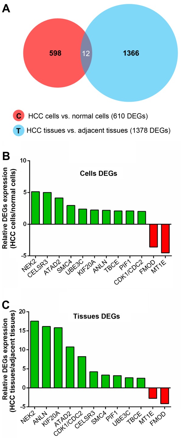
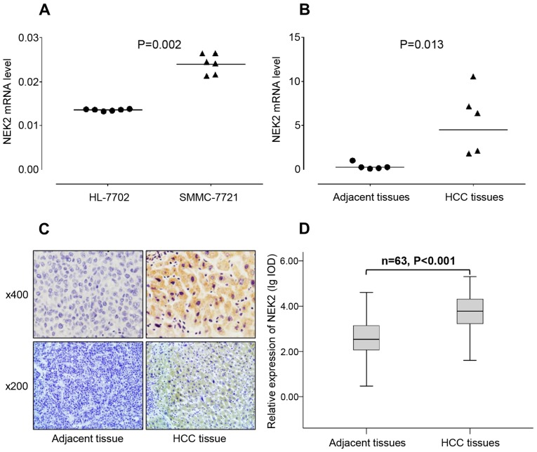
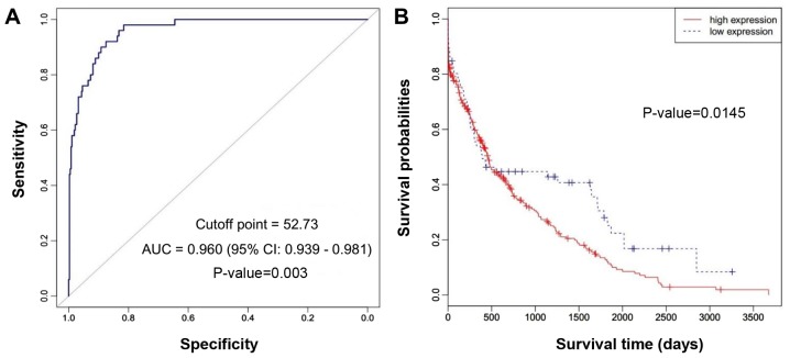
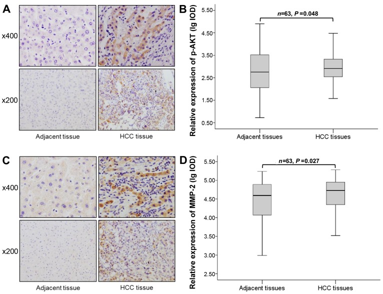
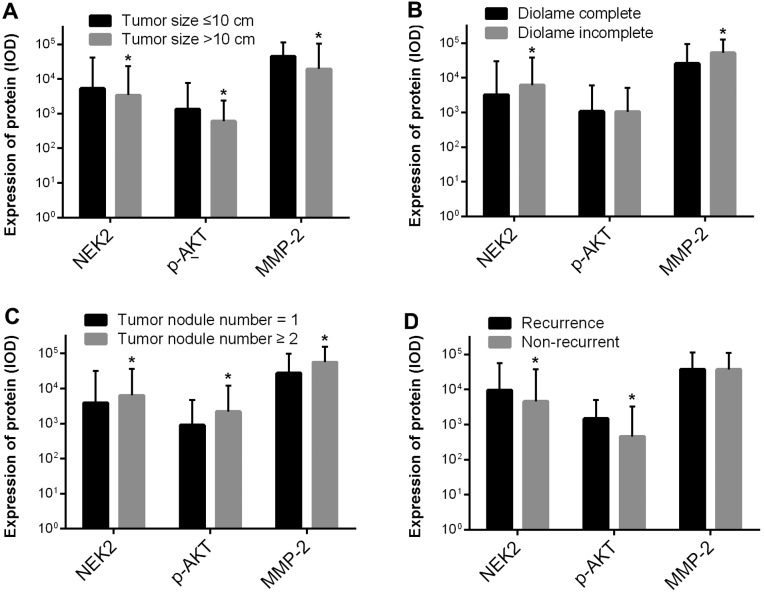
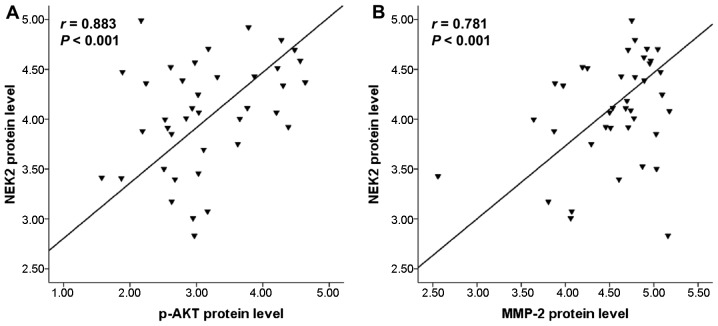
Never in mitosis gene A (NIMA)-related kinase 2 (NEK2) is a microtubule-associated protein that regulates spindle assembly in human cells and is overexpressed in various malignancies. However, the role of NEK2 in hepatocellular carcinoma (HCC) remains undetermined. We performed RNA-seq of the HCC cell line SMMC-7721 and the normal liver cell line HL-7702 using the Ion Proton System. NEK2 expression was detected using quantitative reverse transcription polymerase chain reaction in two cell lines and 5 matched HCC and adjacent non-tumorous liver tissues. The correlation between survival and NEK2 expression was analyzed in 359 patients with HCC using RNASeqV2 data available from The Cancer Genome Atlas (TCGA) website (https://tcga-data.nci.nih.gov/tcga/). The expression of NEK2, phospho-AKT and MMP-2 was evaluated by immunohistochemistry in 63 cases of HCC and matched adjacent non-tumorous liver tissues. Relationships between protein expression and clinicopathological parameters were assessed, and the correlations between NEK2 with phospho-AKT and MMP-2 expressions were evaluated. A total of 610 differentially expressed genes (DEGs) were revealed in the transcriptome comparison, 297 of which were upregulated and 313 were downregulated in HCC. NEK2, as the most obviously different DEG in cells and tissues from the RNA-seq data, was listed as an HCC candidate biomarker for further verification. NEK2 was overexpressed in HCC cells and tissues (P=0.002, P=0.013) and HCC patients with a high expression of NEK2 had a poor prognosis (P=0.0145). Clinical analysis indicated that the overexpression of NEK2 in HCC was significantly correlated with diolame complete (P<0.001), tumor nodule number (P=0.012) and recurrence (P=0.004). NEK2 expression was positively correlated with the expression of phospho-AKT (r=0.883, P<0.01) and MMP-2 (r=0.781, P<0.01). Overexpression of NEK2 was associated with clinicopathological characteristics and poor patient outcomes, suggesting that NEK2 serves as a prognostic biomarker for HCC. Alteration of NEK2 protein levels may contribute to invasion and metastasis of HCC, which may occur through activation of AKT signaling and promotion of MMP-2 expression.
Figures
Similar articles
-
High NEK2 Expression Is a Predictor of Tumor Recurrence in Hepatocellular Carcinoma Patients After Hepatectomy.Anticancer Res. 2016 Feb;36(2):757-62. Anticancer Res. 2016. PMID: 26851035
-
The Prognostic Significance of NEK2 in Hepatocellular Carcinoma: Evidence from a Meta-Analysis and Retrospective Cohort Study.Cell Physiol Biochem. 2018;51(6):2746-2759. doi: 10.1159/000495966. Epub 2018 Dec 12. Cell Physiol Biochem. 2018. PMID: 30562736
-
Low expression of NEK2 is associated with hepatocellular carcinoma progression and poor prognosis.Cancer Biomark. 2017 Jul 19;20(1):101-106. doi: 10.3233/CBM-170586. Cancer Biomark. 2017. PMID: 28759960
-
Prognostic significance of NEK2 in human solid tumors: a systematic review and meta-analysis.Biosci Rep. 2019 Jan 18;39(1):BSR20180618. doi: 10.1042/BSR20180618. Print 2019 Jan 31. Biosci Rep. 2019. PMID: 30578380 Free PMC article.
-
Role of NIMA-related kinase 2 in lung cancer: Mechanisms and therapeutic prospects.Fundam Clin Pharmacol. 2022 Oct;36(5):766-776. doi: 10.1111/fcp.12777. Epub 2022 Apr 2. Fundam Clin Pharmacol. 2022. PMID: 35338518 Review.
Cited by
-
NIMA-related kinase 2 overexpression is associated with poor survival in cancer patients: a systematic review and meta-analysis.Cancer Manag Res. 2019 Jan 3;11:455-465. doi: 10.2147/CMAR.S188347. eCollection 2019. Cancer Manag Res. 2019. PMID: 30655697 Free PMC article.
-
Identification and Validation of Two Lung Adenocarcinoma-Development Characteristic Gene Sets for Diagnosing Lung Adenocarcinoma and Predicting Prognosis.Front Genet. 2020 Dec 21;11:565206. doi: 10.3389/fgene.2020.565206. eCollection 2020. Front Genet. 2020. PMID: 33408736 Free PMC article.
-
Screening Hub Genes as Prognostic Biomarkers of Hepatocellular Carcinoma by Bioinformatics Analysis.Cell Transplant. 2019 Dec;28(1_suppl):76S-86S. doi: 10.1177/0963689719893950. Epub 2019 Dec 11. Cell Transplant. 2019. PMID: 31822116 Free PMC article.
-
Identification of key genes in hepatitis B associated hepatocellular carcinoma based on WGCNA.Infect Agent Cancer. 2021 Mar 16;16(1):18. doi: 10.1186/s13027-021-00357-4. Infect Agent Cancer. 2021. PMID: 33726794 Free PMC article.
-
In Mitosis You Are Not: The NIMA Family of Kinases in Aspergillus, Yeast, and Mammals.Int J Mol Sci. 2022 Apr 6;23(7):4041. doi: 10.3390/ijms23074041. Int J Mol Sci. 2022. PMID: 35409400 Free PMC article. Review.
References
MeSH terms
Substances
LinkOut - more resources
Full Text Sources
Other Literature Sources
Medical
Miscellaneous
