Operative planning aid for optimal endoscopic third ventriculostomy entry points in pediatric cases
- PMID: 28101675
- PMCID: PMC5352741
- DOI: 10.1007/s00381-016-3320-y
Operative planning aid for optimal endoscopic third ventriculostomy entry points in pediatric cases
Abstract
Object: Endoscopic third ventriculostomy (ETV) uses anatomical spaces of the ventricular system to reach the third ventricle floor and create an alternative pathway for cerebrospinal fluid flow. Optimal ETV trajectories have been previously proposed in the literature, designed to grant access to the third ventricle floor without a displacement of eloquent periventricular structures. However, in hydrocephalus, there is a significant variability to the configuration of the ventricular system, implying that the optimal ETV trajectory and cranial entry point needs to be planned on a case-by-case basis. In the current study, we created a mathematical model, which tailors the optimal ETV entry point to the individual case by incorporating the ventricle dimensions.
Methods: We retrospectively reviewed the imaging of 30 consecutive pediatric patients with varying degrees of ventriculomegaly. Three dimensional radioanatomical models were created using preoperative MRI scans to simulate the optimal ETV trajectory and entry point for each case. The surface location of cranial entry points for individual ETV trajectories was recorded as Cartesian coordinates centered at Bregma. The distance from the Bregma in the coronal plane represented as "x", and the distance from the coronal suture in the sagittal plane represented as "y". The correlation between the ventricle dimensions and the x, y coordinates were tested using linear regression models.
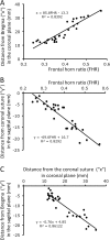
Results: The distance of the optimal ETV entry point from the Bregma in the coronal plane ("x") and from the coronal suture in the sagittal plane ("y") correlated well with the frontal horn ratio (FHR). The coordinates for x and y were fitted along the following linear equations: x = 85.8 FHR-13.3 (r 2 = 0.84, p < 0.001) and y = -69.6 FHR + 16.7 (r 2 = 0.83, p < 0.001).
Conclusion: The surface location of the optimal cranial ETV entry point correlates well with the ventricle size. We provide the first model that can be used as a surgical planning aid for a case specific ETV entry site with the incorporation of the ventricle size.
Keywords: Endoscopic third ventriculostomy; Optimal ETV trajectory; Pediatric hydrocephalus.
Conflict of interest statement
None
Figures


Similar articles
-
Comparative analysis of endoscopic third ventriculostomy trajectories in pediatric cases.J Neurosurg Pediatr. 2015 Dec;16(6):626-32. doi: 10.3171/2015.4.PEDS14430. Epub 2015 Sep 4. J Neurosurg Pediatr. 2015. PMID: 26339953
-
Robot-assisted endoscopic third ventriculostomy: institutional experience in 9 patients.J Neurosurg Pediatr. 2017 Aug;20(2):125-133. doi: 10.3171/2017.3.PEDS16636. Epub 2017 Jun 9. J Neurosurg Pediatr. 2017. PMID: 28598265
-
Single burr hole endoscopic biopsy with third ventriculostomy-measurements and computer-assisted planning.Childs Nerv Syst. 2011 Aug;27(8):1233-41. doi: 10.1007/s00381-011-1405-1. Epub 2011 Feb 16. Childs Nerv Syst. 2011. PMID: 21327590
-
Techniques of endoscopic third ventriculostomy.Neurosurg Clin N Am. 2004 Jan;15(1):51-9. doi: 10.1016/S1042-3680(03)00066-4. Neurosurg Clin N Am. 2004. PMID: 15062403 Review.
-
A pediatric experience with endoscopic third ventriculostomy for hydrocephalus.Can J Neurosci Nurs. 2009;31(2):16-9. Can J Neurosci Nurs. 2009. PMID: 19522457 Review.
References
-
- Amini A, Schmidt RH. Endoscopic third ventriculostomy in a series of 36 adult patients. Neurosurg Focus. 2005;19 - PubMed
MeSH terms
LinkOut - more resources
Full Text Sources
Other Literature Sources
Medical

