. 2017 Jan 19:8:14162.
doi: 10.1038/ncomms14162.

Whole-brain 3D mapping of human neural transplant innervation

Affiliations

Whole-brain 3D mapping of human neural transplant innervation

Jonas Doerr et al. Nat Commun. .

Abstract

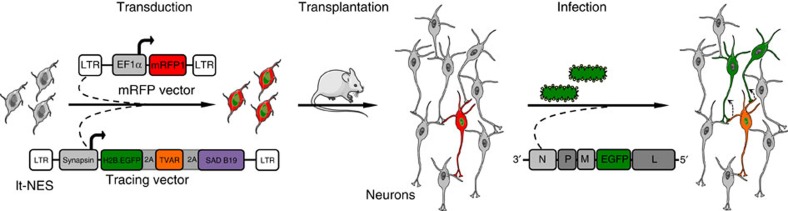
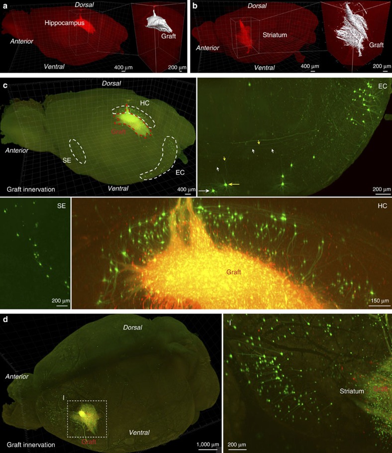
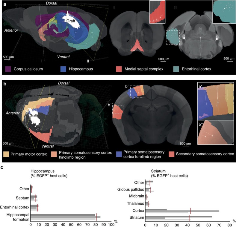
While transplantation represents a key tool for assessing in vivo functionality of neural stem cells and their suitability for neural repair, little is known about the integration of grafted neurons into the host brain circuitry. Rabies virus-based retrograde tracing has developed into a powerful approach for visualizing synaptically connected neurons. Here, we combine this technique with light sheet fluorescence microscopy (LSFM) to visualize transplanted cells and connected host neurons in whole-mouse brain preparations. Combined with co-registration of high-precision three-dimensional magnetic resonance imaging (3D MRI) reference data sets, this approach enables precise anatomical allocation of the host input neurons. Our data show that the same neural donor cell population grafted into different brain regions receives highly orthotopic input. These findings indicate that transplant connectivity is largely dictated by the circuitry of the target region and depict rabies-based transsynaptic tracing and LSFM as efficient tools for comprehensive assessment of host-donor cell innervation.


Conflict of interest statement

O.B. is a co-founder and owns equity of LIFE & BRAIN GmbH. The remaining authors declare no competing financial interests.

Figures

Figure 1
Figure 1. Experimental procedure for tracing host-graft connectivity.
Lt-NES cells were transduced with two lentiviral vectors expressing mRFP1, and an H2B.EGFP fusion protein coupled via 2A peptides to the TVA receptor and the RABV B19 glycoprotein. Ten weeks after transplantation the grafts were infected with RABVΔG-EGFP(EnvA), which enables monosynaptic retrograde tracing of afferent host neurons. Parts of the schematic were produced using Servier Medical Art (http://www.servier.com).
Figure 2
Figure 2. Whole-mount imaging of grafted human lt-NES cells.
(a,b) Human grafts in the hippocampus (a) and striatum (b) as detected by their mRFP1 expression. Insets depict magnified graft cores as determined by 3D surface rendering. (c,d) Co-visualization of mRFP1 and EGFP fluorescence identifies RABVΔG-EGFP-infected donor cells while exclusively EGFP+ cells represent retrogradely labelled host neurons connected to engrafted neurons. (c) Hippocampal grafts (HC) receive orthotopic input from the entorhinal cortex (EC; coloured arrows indicate neurons and their corresponding axon), the septum (SE) and intrahippocampal host neurons within the pyramidal cell layer and stratum oriens. Striatal grafts (d) are largely innervated by cortical neurons as well as adjacent striatal neurons. Shown are representative images from n=3 cleared brains per transplantation site; scale bars as indicated.
Figure 3
Figure 3. Anatomical distribution of host neurons projecting onto grafted human neurons.
Simultaneous representation of LSFM-recorded EGFP-labelled neurons and co-registration with MRI data in recipient mouse brains with (a) hippocampal and (b) striatal grafts. Host cells innervating the hippocampal graft are allocated to adjacent hippocampal territories, septum and entorhinal cortex. In brains harbouring striatal grafts, MRI co-registration permitted the allocation of host input neurons to cortical subregions such as motor cortex, secondary somatosensory cortex and distinct areas within primary somatosensory cortex. Shown are representative images from n=3 cleared brains per transplantation site; scale bars as indicated. (c) Quantification and anatomical allocation of EGFP+ host neurons innervating the grafted human neurons. Red vertical bars indicate mean values for 3 recipient animals each (grey horizontal bars). Shown is the percentage of all detected EGFP+ host neurons.

References

    1. Thompson L. H. & Björklund A. Reconstruction of brain circuitry by neural transplants generated from pluripotent stem cells. Neurobiol. Dis. 79, 28–40 (2015). - PubMed
    1. Steinbeck J. & Studer L. Moving stem cells to the clinic: potential and limitations for brain repair. Neuron 86, 187–206 (2015). - PMC - PubMed
    1. Debanne D. et al.. Paired-recordings from synaptically coupled cortical and hippocampal neurons in acute and cultured brain slices. Nat. Protoc. 3, 1559–1568 (2008). - PubMed
    1. Svoboda K., Denk W., Kleinfeld D. & Tank D. W. In vivo dendritic calcium dynamics in neocortical pyramidal neurons. Nature 385, 161–165 (1997). - PubMed
    1. Antic S. D., Empson R. M. & Knopfel T. Voltage imaging to understand connections and functions of neuronal circuits. J. Neurophysiol. 116, 135–152 (2016). - PMC - PubMed

Publication types

LinkOut - more resources