. 2017 Feb 21;56(7):957-970.
doi: 10.1021/acs.biochem.6b00888. Epub 2017 Feb 6.

Structural Analysis of the Glycosylated Intact HIV-1 gp120-b12 Antibody Complex Using Hydroxyl Radical Protein Footprinting

Affiliations

Structural Analysis of the Glycosylated Intact HIV-1 gp120-b12 Antibody Complex Using Hydroxyl Radical Protein Footprinting

Xiaoyan Li et al. Biochemistry. .

Abstract

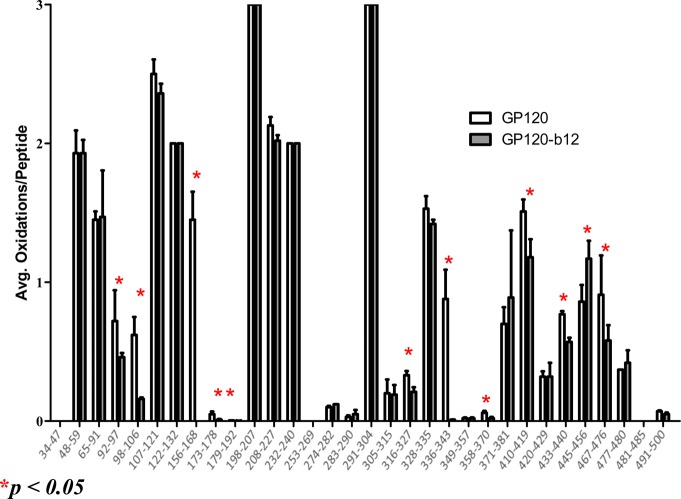
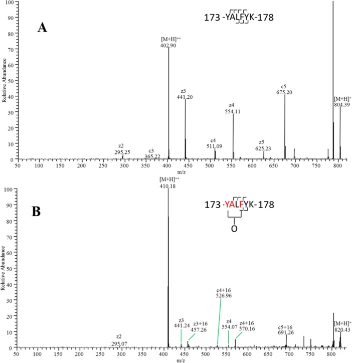
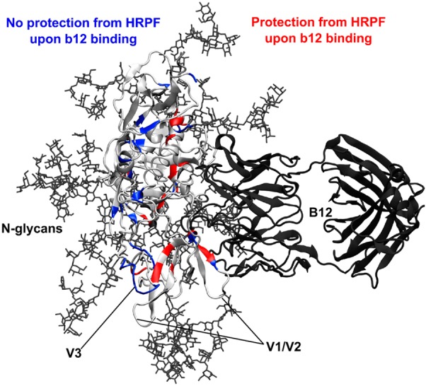
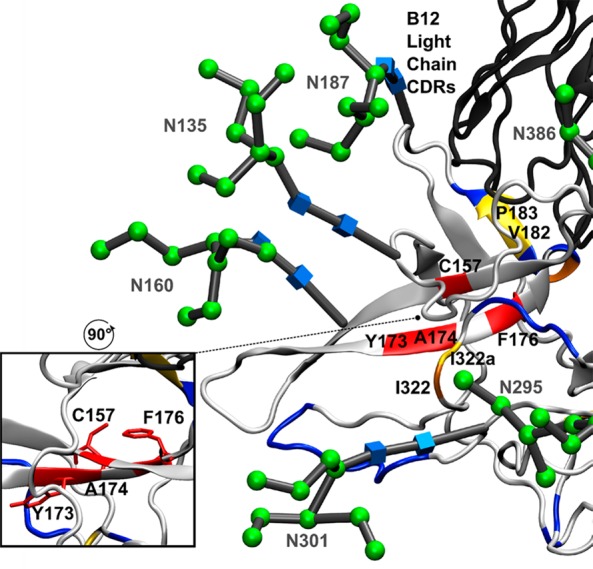
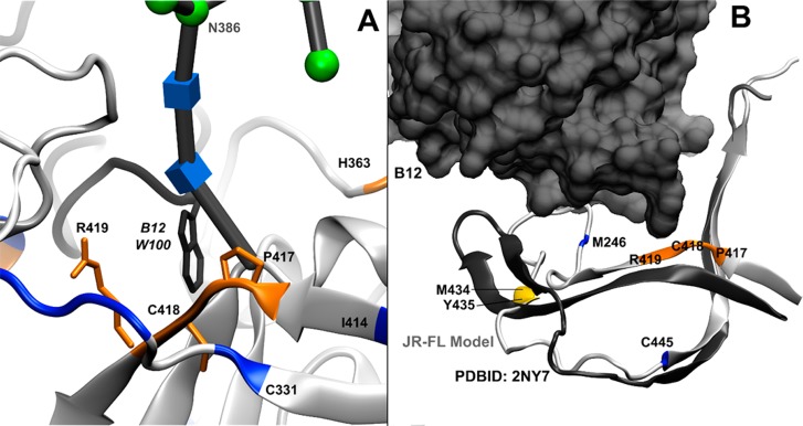
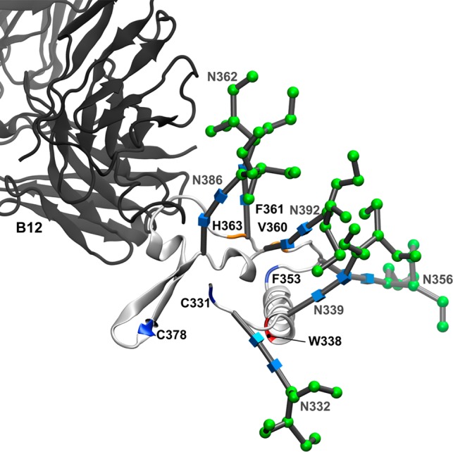
Glycoprotein gp120 is a surface antigen and virulence factor of human immunodeficiency virus 1. Broadly neutralizing antibodies (bNAbs) that react to gp120 from a variety of HIV isolates offer hope for the development of broadly effective immunogens for vaccination purposes, if the interactions between gp120 and bNAbs can be understood. From a structural perspective, gp120 is a particularly difficult system because of its size, the presence of multiple flexible regions, and the large amount of glycosylation, all of which are important in gp120-bNAb interactions. Here, the interaction of full-length, glycosylated gp120 with bNAb b12 is probed using high-resolution hydroxyl radical protein footprinting (HR-HRPF) by fast photochemical oxidation of proteins. HR-HRPF allows for the measurement of changes in the average solvent accessible surface area of multiple amino acids without the need for measures that might alter the protein conformation, such as mutagenesis. HR-HRPF of the gp120-b12 complex coupled with computational modeling shows a novel extensive interaction of the V1/V2 domain, probably with the light chain of b12. Our data also reveal HR-HRPF protection in the C3 domain caused by interaction of the N330 glycan with the b12 light chain. In addition to providing information about the interactions of full-length, glycosylated gp120 with b12, this work serves as a template for the structural interrogation of full-length glycosylated gp120 with other bNAbs to better characterize the interactions that drive the broad specificity of the bNAb.


Conflict of interest statement

The authors declare the following competing financial interest(s): J.S.S. discloses a significant ownership share of Photochem Technologies, LLC, a small company active in the area of hydroxyl radical protein footprinting.

Figures

Figure 1
Figure 1
Sequence of HIV-1 gp120 (JR-FL). The sequences covered by HR-HRPF experiments are overlined with green. The identified oxidation sites are shown in red text with protected sites underlined and the sole exposed site in italics. The heavily occupied N-linked glycosylation sites are shown in green text. The variable domains are labeled above the sequence.
Figure 2
Figure 2
Peptide level HRPF of gp120 footprinting for gp120 alone (white) and the gp120–b12 complex (gray) (mean ± standard deviation; n = 3). Peptides highlighted with a red star showed a statistically significant change in oxidation extent upon gp120–b12 binding (α ≤ 0.05).
Figure 3
Figure 3
ETD spectrum of peptide 143-YALFYK-148 and its oxidation products. (A) Unoxidized peptide 173-YALFYK-178. (B) Mixture of singly oxidized isomers. By measuring the ratio of oxidized product ions to total product ions for each fragment, oxidation can be quantified to occur on F176 and the Y173-A174 fragment (on the basis of CID data and the relative reactivities of alanine and tyrosine, almost no oxidation occurs on A174).
Figure 4
Figure 4
Glycosylation and HRPF of gp120. A model of full-length glycosylated gp120 (ribbon protein in light gray, licorice Man9GlcNAc2 glycans in dark gray) was generated and relaxed by MD simulation and then aligned with the crystal structure of stabilized gp120 bound to the b12 Fab (aligned gp120 not shown, ribbon b12 in dark gray) (PDB entry 2NY7). Residues that showed protection from HRPF upon b12 binding are colored red. Residues that showed no protection from HRPF are colored blue. Residue C445 (also blue) showed an exposure to HRPF upon b12.
Figure 5
Figure 5
HR-HRPF and glycosylation of the V1/V2 and V3 domains. Aligned model of full-length glycosylated gp120 after MD simulation (light gray ribbon) bound to the b12 Fab (dark gray ribbon) (PDB entry 2NY7). Residues that showed >80% protection from HR-HRPF upon b12 binding are colored red. Residues that showed between 40 and 80% protection from HR-HRPF upon b12 binding are colored orange. Residues that showed statistically significant protection from HR-HRPF of <40% are colored yellow. Residues showing no protection from HR-HRPF upon b12 binding are colored blue. The CDR loops of the light chain modeled to potentially interact with the V1/V2 domain are shown. The inset shows the orientation of the C157, F176, and Y173 side chains relative to the β-sheet.
Figure 6
Figure 6
HR-HRPF protection suggests stabilization of the helix α1–helix α5 interaction upon binding of b12 to helix α5. Helix α1 of the C1 domain (green) shows protection of four residues: M95 and W96 in the loop at the N-terminus of helix α1 and M99 and M104 within helix α1. The sole residue probed in helix α5, M475, interacts directly with a CDR loop of the b12 heavy chain (dark gray), with helix α5 (light gray) packing against the N-terminus of helix α1. Residues that showed >80% protection from HR-HRPF upon b12 binding are colored red. Residues that showed between 40 and 80% protection from HR-HRPF upon b12 binding are colored orange. Residues that showed statistically significant protection from HR-HRPF of <40% are colored yellow. Residues showing no protection from HR-HRPF upon b12 binding are colored blue.
Figure 7
Figure 7
b12 both interacts directly with domain C4 and alters its dynamics. Residues that showed >80% protection from HR-HRPF upon b12 binding are colored red. Residues that showed between 40 and 80% protection from HR-HRPF upon b12 binding are colored orange. Residues that showed statistically significant protection from HR-HRPF of <40% are colored yellow. Residues showing no protection from HR-HRPF upon b12 binding are colored blue. Residue C445 (also blue) showed an increase in the level of oxidation upon b12 binding. (A) P417, C418, and R419 of strand β19 (orange ribbon and licorice) interact directly with W100 (dark gray ribbon and licorice) of the CDR H3 loop of the b12 heavy chain, which extends out from the bulk of the b12 IgG fold. (B) Model of full-length, glycosylated gp120 (light gray) aligned with the crystal structure of stabilized, b12-bound gp120 (dark gray), with only the C4 domain backbone shown for the sake of clarity and the b12 antibody shown as a space-filling model. Strand β22/β23 remains largely unperturbed; however, strands β20 and β21 are much more flexible in the full-length, glycosylated, unbound gp120 MD simulation. HR-HRPF data of M426 show no protection upon b12 binding, suggesting that there is no appreciable change in the conformation or dynamics of this residue in strand β20. However, the protection of M434 and Y435 suggests that strand β21 does experience a stabilization upon b12 binding.
Figure 8
Figure 8
Interaction of b12 with the C3 domain of gp120 mediated by N-linked glycosylation. Backbone and N-linked glycans of the C3 domain from the full-length, glycosylated gp120 MD simulation (light gray ribbon) aligned with the b12 Fab fragment (dark gray ribbon). All other gp120 domains have been excluded for the sake of clarity. Residues that showed >80% protection from HR-HRPF upon b12 binding are colored red. Residues that showed between 40 and 80% protection from HR-HRPF upon b12 binding are colored orange. Residues showing no protection from HR-HRPF upon b12 binding are colored blue. Glycans are labeled and shown as 3D-SNFG symbols, which are positioned at each residue’s ring center.

Similar articles

Cited by

References

    1. Kowalski M.; Potz J.; Basiripour L.; Dorfman T.; Goh W. C.; Terwilliger E.; Dayton A.; Rosen C.; Haseltine W.; Sodroski J. (1987) Functional regions of the envelope glycoprotein of human immunodeficiency virus type 1. Science 237, 1351–1355. 10.1126/science.3629244. - DOI - PubMed
    1. Nara P. L.; Garrity R. R.; Goudsmit J. (1991) Neutralization of HIV-1: a paradox of humoral proportions. FASEB J. 5, 2437–2455. - PubMed
    1. Lasky L. A.; Groopman J. E.; Fennie C. W.; Benz P. M.; Capon D. J.; Dowbenko D. J.; Nakamura G. R.; Nunes W. M.; Renz M. E.; Berman P. W. (1986) Neutralization of the AIDS retrovirus by antibodies to a recombinant envelope glycoprotein. Science 233, 209–212. 10.1126/science.3014647. - DOI - PubMed
    1. Kwong P. D.; Wyatt R.; Robinson J.; Sweet R. W.; Sodroski J.; Hendrickson W. A. (1998) Structure of an HIV gp120 envelope glycoprotein in complex with the CD4 receptor and a neutralizing human antibody. Nature 393, 648–659. 10.1038/31405. - DOI - PMC - PubMed
    1. Wyatt R.; Kwong P. D.; Desjardins E.; Sweet R. W.; Robinson J.; Hendrickson W. A.; Sodroski J. G. (1998) The antigenic structure of the HIV gp120 envelope glycoprotein. Nature 393, 705–711. 10.1038/31514. - DOI - PubMed

Publication types

MeSH terms