

Soluble P-selectin rescues mice from anthrax lethal toxin-induced mortality through PSGL-1 pathway-mediated correction of hemostasis
- PMID: 28102766
- PMCID: PMC5711406
- DOI: 10.1080/21505594.2017.1282027
Soluble P-selectin rescues mice from anthrax lethal toxin-induced mortality through PSGL-1 pathway-mediated correction of hemostasis
Abstract
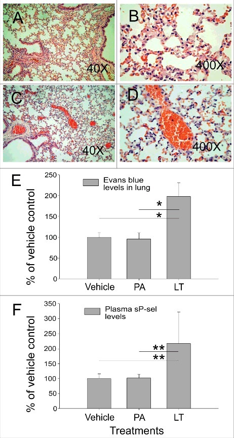
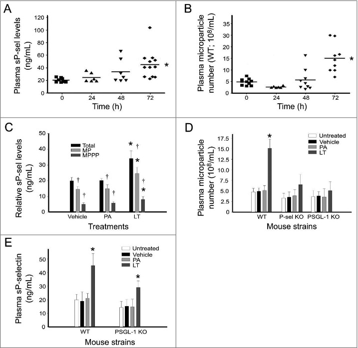
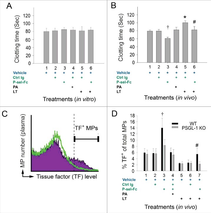
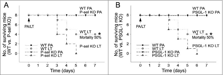
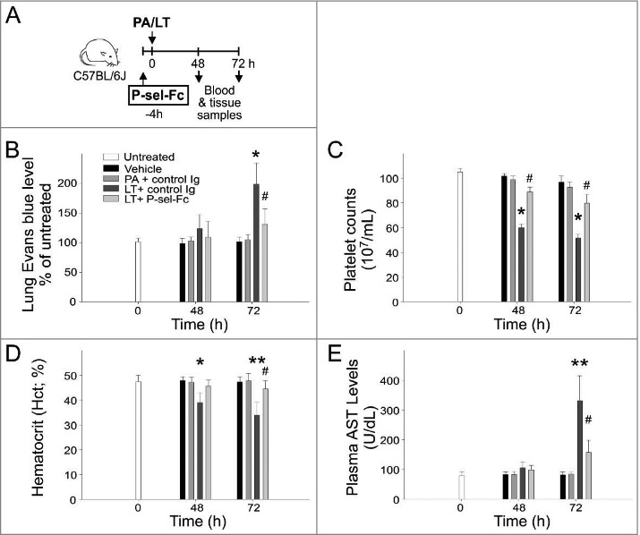
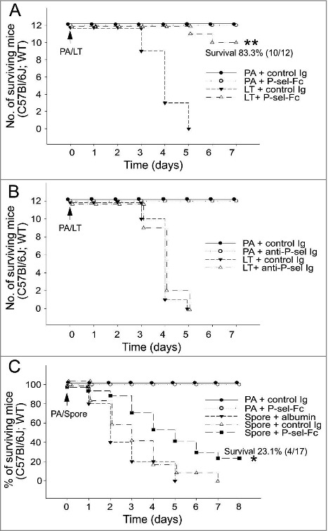
As one of the virulence factors of Bacillus anthracis, lethal toxin (LT) induces various pathogenic responses including the suppression of the coagulation system. In this study, we observed that LT markedly increased the circulating soluble P-selectin (sP-sel) levels and microparticle (MP) count in wild-type but not P-selectin (P-sel, Selp-/-) or P-sel ligand-1 (PSGL-1, Selplg-/-) knockout mice. Because sP-sel induces a hypercoagulable state through PSGL-1 pathway to generate tissue factor-positive MPs, we hypothesized that the increase in plasma sP-sel levels can be a self-rescue response in hosts against the LT-mediated suppression of the coagulation system. In agreement with our hypothesis, our results indicated that compared with wild-type mice, Selp-/- and Selplg-/- mice were more sensitive to LT. In addition, the recombinant sP-sel treatment markedly ameliorated LT-mediated pathogenesis and reduced mortality. As a result, elicitation of circulating sP-sel is potentially a self-rescue response, which is beneficial to host recovery from an LT-induced hypocoagulation state. These results suggest that the administration of sP-sel is likely to be useful in the development of a new strategy to treat anthrax.
Keywords: P-selectin ligand 1; Soluble P-selectin; anthrax lethal toxin; hemostasis; microparticle.
Figures
Comment in
-
Self-defense against Bacillus anthracis toxins: Is P-selectin the key?Virulence. 2017 Oct 3;8(7):1059-1061. doi: 10.1080/21505594.2017.1304344. Epub 2017 Mar 10. Virulence. 2017. PMID: 28281892 Free PMC article. No abstract available.
Similar articles
-
Soluble P-selectin rescues viper venom-induced mortality through anti-inflammatory properties and PSGL-1 pathway-mediated correction of hemostasis.Sci Rep. 2016 Oct 25;6:35868. doi: 10.1038/srep35868. Sci Rep. 2016. PMID: 27779216 Free PMC article.
-
Acquired coagulant factor VIII deficiency induced by Bacillus anthracis lethal toxin in mice.Virulence. 2015;6(5):466-75. doi: 10.1080/21505594.2015.1031454. Epub 2015 Apr 23. Virulence. 2015. PMID: 25906166 Free PMC article.
-
Mouse susceptibility to anthrax lethal toxin is influenced by genetic factors in addition to those controlling macrophage sensitivity.Infect Immun. 2004 Aug;72(8):4439-47. doi: 10.1128/IAI.72.8.4439-4447.2004. Infect Immun. 2004. PMID: 15271901 Free PMC article.
-
New insights into the functions of anthrax toxin.Expert Rev Mol Med. 2006 Apr 11;8(7):1-18. doi: 10.1017/S1462399406010714. Expert Rev Mol Med. 2006. PMID: 16608555 Review.
-
The P-selectin, tissue factor, coagulation triad.J Thromb Haemost. 2005 Aug;3(8):1590-6. doi: 10.1111/j.1538-7836.2005.01373.x. J Thromb Haemost. 2005. PMID: 16102023 Review.
Cited by
-
Dual phenotypic characteristics of P-selectin in a mouse model of hemorrhagic shock and hepatectomy.Heliyon. 2023 Jul 28;9(8):e18627. doi: 10.1016/j.heliyon.2023.e18627. eCollection 2023 Aug. Heliyon. 2023. PMID: 37554775 Free PMC article.
-
Opportunistic gill infection is associated with TiO2 nanoparticle-induced mortality in zebrafish.PLoS One. 2021 Jul 20;16(7):e0247859. doi: 10.1371/journal.pone.0247859. eCollection 2021. PLoS One. 2021. PMID: 34283836 Free PMC article.
-
Exposure to Dengue Envelope Protein Domain III Induces Nlrp3 Inflammasome-Dependent Endothelial Dysfunction and Hemorrhage in Mice.Front Immunol. 2021 Feb 25;12:617251. doi: 10.3389/fimmu.2021.617251. eCollection 2021. Front Immunol. 2021. PMID: 33717109 Free PMC article.
-
Simulation of Hemorrhage Pathogenesis in Mice through Dual Stimulation with Dengue Envelope Protein Domain III-Coated Nanoparticles and Antiplatelet Antibody.Int J Mol Sci. 2023 May 25;24(11):9270. doi: 10.3390/ijms24119270. Int J Mol Sci. 2023. PMID: 37298220 Free PMC article.
-
Thioacetamide-induced liver damage and thrombocytopenia is associated with induction of antiplatelet autoantibody in mice.Sci Rep. 2019 Nov 25;9(1):17497. doi: 10.1038/s41598-019-53977-7. Sci Rep. 2019. PMID: 31767905 Free PMC article.
References
-
- Friebe S, van der Goot FG, Burgi J. The ins and outs of anthrax toxin. Toxins (Basel) 2016; 8:e69; PMID:26978402; http://dx.doi.org/10.3390/toxins8030069 - DOI - PMC - PubMed
-
- Lowe DE, Glomski IJ. Cellular and physiological effects of anthrax exotoxin and its relevance to disease. Front Cell Infect Microbiol 2012; 2:76; PMID:22919667; http://dx.doi.org/10.3389/fcimb.2012.00076 - DOI - PMC - PubMed
-
- Kau JH, Shih YL, Lien TS, Lee CC, Huang HH, Lin HC, Sun DS, Chang HH. Activated protein C ameliorates Bacillus anthracis lethal toxin-induced lethal pathogenesis in rats. J Biomed Sci 2012; 19:98; PMID:23170801; http://dx.doi.org/10.1186/1423-0127-19-98 - DOI - PMC - PubMed
-
- Kau JH, Sun DS, Huang HH, Wong MS, Lin HC, Chang HH. Role of visible light-activated photocatalyst on the reduction of anthrax spore-induced mortality in mice. PloS One 2009; 4:e4167; PMID:19132100; http://dx.doi.org/10.1371/journal.pone.0004167 - DOI - PMC - PubMed
-
- Kau JH, Sun DS, Tsai WJ, Shyu HF, Huang HH, Lin HC, Chang HH. Antiplatelet activities of anthrax lethal toxin are associated with suppressed p42/44 and p38 mitogen-activated protein kinase pathways in the platelets. J Infect Dis 2005; 192:1465-74; PMID:16170766; http://dx.doi.org/10.1086/491477 - DOI - PubMed
Publication types
MeSH terms
Substances
LinkOut - more resources
Full Text Sources
Other Literature Sources
Medical
Research Materials
Miscellaneous