. 2017 Mar 4;13(3):473-485.
doi: 10.1080/15548627.2016.1274596. Epub 2017 Jan 19.

PARK2-dependent mitophagy induced by acidic postconditioning protects against focal cerebral ischemia and extends the reperfusion window

Affiliations

PARK2-dependent mitophagy induced by acidic postconditioning protects against focal cerebral ischemia and extends the reperfusion window

Zhe Shen et al. Autophagy. .

Abstract

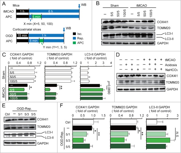
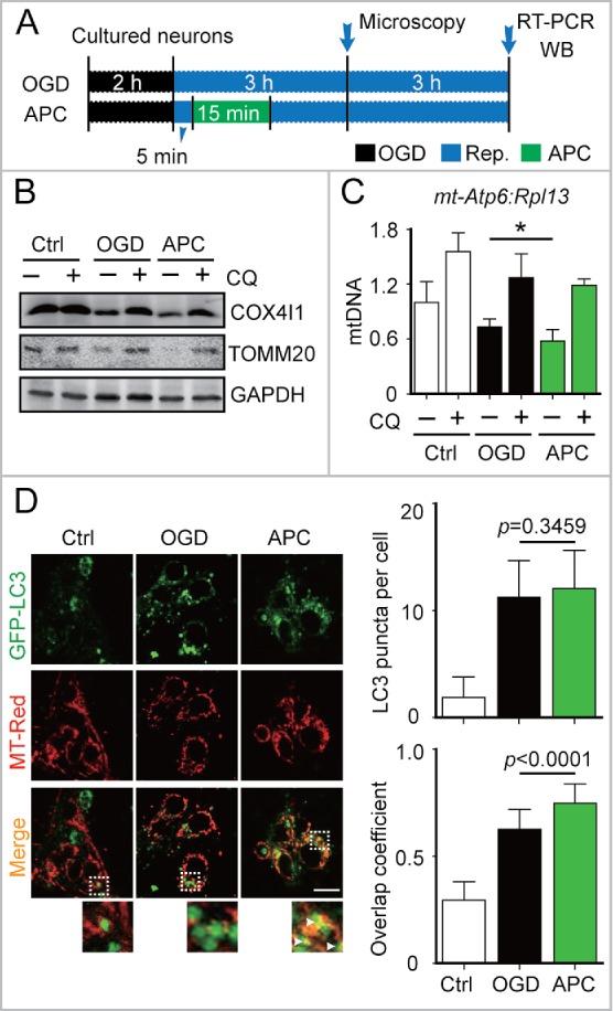
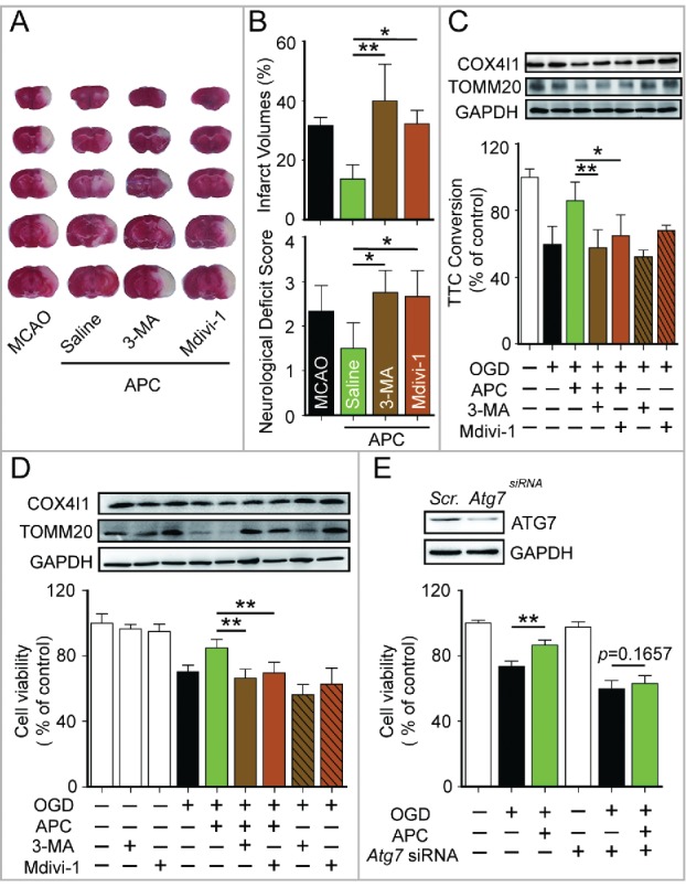
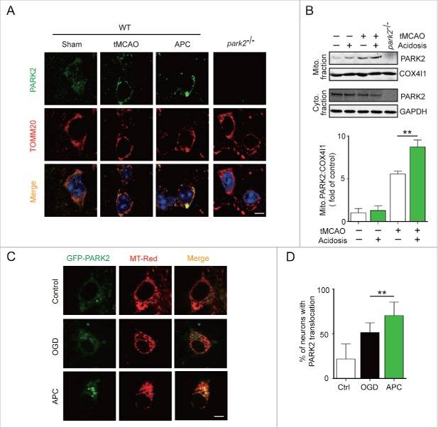
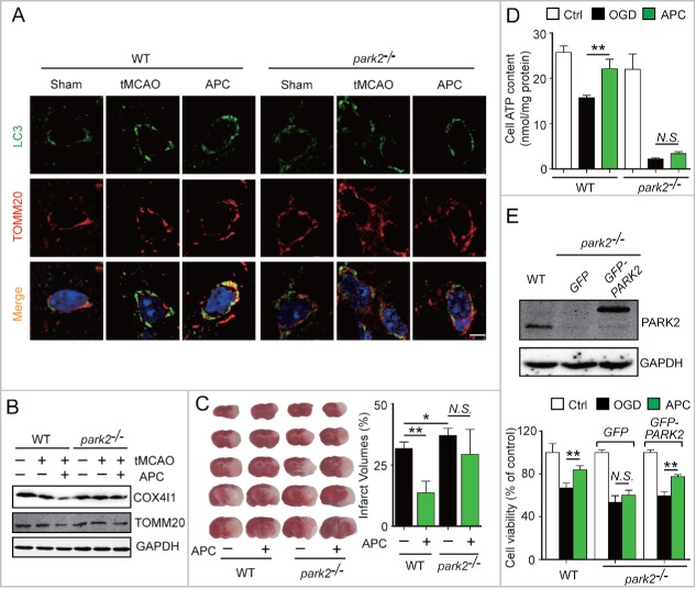
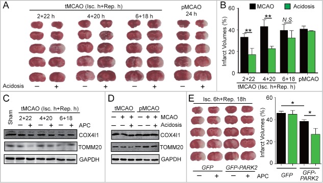
Prompt reperfusion after cerebral ischemia is critical for neuronal survival. Any strategies that extend the limited reperfusion window will be of great importance. Acidic postconditioning (APC) is a mild acidosis treatment that involves inhaling CO2 during reperfusion following ischemia. APC attenuates ischemic brain injury although the underlying mechanisms have not been elucidated. Here we report that APC reinforces ischemia-reperfusion-induced mitophagy in middle cortical artery occlusion (MCAO)-treated mice, and in oxygen-glucose deprivation (OGD)-treated brain slices and neurons. Inhibition of mitophagy compromises neuroprotection conferred by APC. Furthermore, mitophagy and neuroprotection are abolished in Park2 knockout mice, indicating that APC-induced mitophagy is facilitated by the recruitment of PARK2 to mitochondria. Importantly, in MCAO mice, APC treatment extended the effective reperfusion window from 2 to 4 h, and this window was further extended to 6 h by exogenously expressing PARK2. Taken together, we found that PARK2-dependent APC-induced mitophagy renders the brain resistant to ischemic injury. APC treatment could be a favorable strategy to extend the thrombolytic time window for stroke therapy.

Keywords: PARK2; acidic postconditioning; cerebral ischemia; mitophagy; neuroprotection; time window.


Figures

Figure 1.
Figure 1.
Acidic postconditioning activates mitophagy after cerebral ischemia in vivo and in vitro. (A) The schematic protocol of surgery for cerebral ischemia with or without acidic postconditioning (APC). For the in vivo model, mice were subjected to 1-h MCAO and then treated by inhaling 20% CO2 for 5 min at 5, 50, or 100 min after reperfusion. X indicates the duration of reperfusion. Mice of sham groups inhaled CO2 at corresponding times. For the in vitro model, OGD-treated corticostriatal slices, slices were subjected to OGD for 15 min and then treated with acidic buffer (pH 6.8) for 1, 3, or 5 min at 5 min after the onset of reperfusion. Y indicates the duration of APC treatment. (B) and C) COX4I1, TOMM20 and LC3 protein levels were determined by western blot analysis at 6 after the onset of reperfusion in tMCAO mice. (D) NaHCO3 was infused at 3 min before APC treatment (inhaling 20% CO2), which was applied for 5 min at 5 min after reperfusion of MCAO. After 6 h of reperfusion, COX4I1 and TOMM20 protein levels were determined by western blot analysis. NaHCO3 administration reversed APC-induced mitochondrial mass reduction. ((E)and F) COX4I1, TOMM20 and LC3 protein levels were determined by western blot analysis at 1 h after the onset of reperfusion in vitro. Data are expressed as mean ± SD n = 3 for each group. *P < 0.05 and **P < 0.01 vs. the indicated groups. N.S., not statistically significant.
Figure 2.
Figure 2.
APC-activated mitophagy in primary cultured neurons. (A) OGD and APC protocols in primary cultured neurons. Primary neuronal cultures were subjected to OGD for 2 h and treated with acidosis for 15 min at 5 min after the onset of reperfusion. (B) After 6 h of reperfusion, TOMM20 and COX4I1 protein levels were determined by western blot analysis in the presence or absence of chloroquine (CQ), and (C) relative mitochondrial DNA levels as indicated by the ratio of mt-Atp6 (mitochondria-encoded DNA) to Rpl13 (nucleus encoded DNA) were assessed by real-time PCR. (D) Cells transfected with AAV-GFP-LC3 were loaded with MitoTracker Red (MT-Red; 100 nmol/L). Images were captured at 3 h after the onset of reperfusion by confocal microscopy. Areas in the white boxes are enlarged at the bottom. Right upper panel columns represent the numbers of GFP-LC3-positive puncta per cell, and the lower panel columns represent Manders' overlap coefficient. At least 30 cells from 3 independent experiments for each group were included. Scale bar: 20 µm. Data are expressed as mean ± SD n = 3 for each group. *P < 0.05 vs. the indicated groups.
Figure 3.
Figure 3.
Mitophagy inhibition blocks APC-mediated neuroprotection. (A) and B) Mice were subjected to 1-h MCAO and then treated by inhaling 20% CO2 for 5 min at 5 min after reperfusion. (A) Representative brain slices after TTC staining are shown. (B) Infarct volumes and neurological deficit scores were measured 24 h after surgery. 7.5 µg of 3-methyladenine (3-MA, intracerebroventricular) or 3 mg/kg mdivi-1 (intraperitoneal) was injected at the onset of reperfusion. n = 4 to 6 for each group. Corticostriatal slices (C) and cultured neurons (D) were subjected to OGD and then treated with acidic buffer (pH 6.8) for 5 or 15 min at 5 min after the onset of reperfusion. The inhibitors, 1.25 mmol/L 3-MA or 25 µmol/L mdivi-1, were applied at the onset of reperfusion. COX4I1 and TOMM20 levels were examined by western blot at 1 or 6 h after the onset of reperfusion respectively (upper panel). Corticostriatal slice viability was quantified by TTC assay at 1 h after reperfusion, and cell viability was assessed by MTT assay at 24 h after reperfusion. (E) Atg7 was knocked down by transfection with microRNA 72 h before OGD. The knockdown of Atg7 expression was confirmed by western blot (upper panel). A scrambled siRNA sequence was used as a negative control (scr.). Cell viability was assessed by MTT assay at 24 h after reperfusion. APC failed to improve cell viability in Atg7 knockdown neurons. n = 3 for each group. Data are expressed as mean ± SD. *P < 0.05 and **P < 0.01 vs. the indicated groups.
Figure 4.
Figure 4.
APC facilitates PARK2 translocation onto mitochondria. Wild-type or park2−/− mice were subjected to 1-h MCAO and then treated by inhaling 20% CO2 for 5 min at 5 min after reperfusion. (A) APC-induced PARK2 translocation onto mitochondria can be seen at 2 h after the onset of reperfusion (green, upper panels). Mitochondria are labeled with the mitochondrial marker TOMM20 (red, lower panels). Nuclei were stained with DAPI. (B) PARK2 levels in both mitochondrial (mito.) and cytosolic (cyto.) fractions were determined by western blot analysis at 2 h after the onset of reperfusion. n = 3 for each group. (C) and D) Primary cultured neurons were transfected with AAV-GFP-PARK2 in advance of treatment. They were then subjected to 2-h OGD and treated with acidic buffer for 15 min at 5 min after the onset of reperfusion. Cells were loaded with MitoTracker Red (MT-Red), and images were captured by confocal microscopy. (C) Images show representative examples from 3 independent experiments. Scale bar: 20 µm. (D) Columns represent the percentage of neurons with PARK2 translocation onto mitochondria. At least 100 cells from 3 independent experiments for each group were included. Data are expressed as mean ± SD. ** P < 0.01 vs. the indicated groups.
Figure 5.
Figure 5.
PARK2 is required for APC-induced activation of mitophagy and neuroprotection. Wild-type or park2−/− mice were subjected to 1-h MCAO and then treated by inhaling 20% CO2 for 5 min at 5 min after reperfusion. (A) APC-activated mitophagy at 3 h after the onset of reperfusion was visualized using the autophagosome marker LC3 (green, upper panels) and the mitochondrial marker TOMM20 (red, lower panels). Nuclei were stained with DAPI. (B) COX4I1 and TOMM20 levels were determined by western blot at 6 h after the onset of reperfusion. n = 3 for each group. (C) Infarct volumes were quantified by TTC staining at 24 h after surgery. n = 6 for each group. (D) Primary cultured neurons were subjected to 2-h OGD, and treated with acidosis for 15 min at 5 min after the onset of reperfusion. ATP levels in cellular supernatants were determined at 24 h after the onset of reperfusion. n = 3 for each group. (E) Primary cultured neurons were transfected with AAV-GFP-PARK2 in advance. The neurons were then treated as described in (D). The PARK2 level in wild-type, park2−/− and park2−/− with GFP-PARK2 neurons was measured by western blot (left panel). Cell viability was examined by MTT assay after 24 h of reperfusion (right panel). n = 3 for each group. Data are expressed as mean ± SD. * P < 0.05 and ** P < 0.01 vs. the indicated groups. N.S., not statistically significant.
Figure 6.
Figure 6.
APC-activated mitophagy extends the reperfusion window for cerebral ischemia. (A)and B) Mice were subjected to permanent middle cerebral artery occlusion (pMCAO) for 24 h or transient middle cerebral artery occlusion (tMCAO) for the indicated durations of reperfusion. Mice with acidosis treatment were treated by inhaling 20% CO2 for 5 min at 1 h after the onset of occlusion (pMCAO) or at 5 min after reperfusion (tMCAO), respectively. (A) A representative TTC-stained brain slice from each group is shown. (B) Infarct volumes were determined 24 h after surgery by TTC staining. n = 6 for each group. (C) and D) The impact of APC on the expression of COX4I1 and TOMM20 in ipsilateral cortical tissue after (C) the indicated tMCAO procedures; (D) tMCAO treatment with 1 h of ischemia + 5 h of reperfusion compared with pMCAO treatment with 6 h of ischemia. (E) Mice were injected with AAV-GFP-PARK2 one month in advance of treatment. They were then subjected to 6-h MCAO and then treated by inhaling 20% CO2 for 5 min at 5 min after reperfusion. Infarct volumes were determined by TTC staining 24 h after surgery, and a representative TTC-stained brain slice from each group is shown. n = 6 for each group. Data are expressed as mean ± SD. * P < 0.05 and ** P < 0.01 vs. the indicated groups. N.S., not statistically significant.

Similar articles

Cited by

References

    1. Moskowitz MA. Brain protection: maybe yes, maybe no. Stroke 2010; 41:S85-6; PMID:20876513; http://dx.doi.org/10.1161/STROKEAHA.110.598458 - DOI - PubMed
    1. Del Zoppo GJ, Saver JL, Jauch EC, Adams HP Jr, American Heart Association Stroke C . Expansion of the time window for treatment of acute ischemic stroke with intravenous tissue plasminogen activator: a science advisory from the American Heart Association/American Stroke Association. Stroke 2009; 40:2945-8; PMID:19478221; http://dx.doi.org/10.1161/STROKEAHA.109.192535 - DOI - PMC - PubMed
    1. Zhao H, Sapolsky RM, Steinberg GK. Interrupting reperfusion as a stroke therapy: ischemic postconditioning reduces infarct size after focal ischemia in rats. J Cereb Blood Flow Metab 2006; 26:1114-21; PMID:16736038; http://dx.doi.org/10.1038/sj.jcbfm.9600233 - DOI - PubMed
    1. Leconte C, Tixier E, Freret T, Toutain J, Saulnier R, Boulouard M, Roussel S, Schumann-Bard P, Bernaudin M. Delayed hypoxic postconditioning protects against cerebral ischemia in the mouse. Stroke 2009; 40:3349-55; PMID:19628803; http://dx.doi.org/10.1161/STROKEAHA.109.557314 - DOI - PubMed
    1. Fan YY, Zhang XN, He P, Shen Z, Shen Y, Wang XF, Hu WW, Chen Z. Transient lack of glucose but not O2 is involved in ischemic postconditioning-induced neuroprotection. CNS Neuroscience Therapeutics 2013; 19:30-7; http://dx.doi.org/10.1111/cns.12033 - DOI - PMC - PubMed

MeSH terms