. 2017 Jan 19;13(1):e1006543.
doi: 10.1371/journal.pgen.1006543. eCollection 2017 Jan.

Protein Phosphatase 1 Down Regulates ZYG-1 Levels to Limit Centriole Duplication

Affiliations

Protein Phosphatase 1 Down Regulates ZYG-1 Levels to Limit Centriole Duplication

Nina Peel et al. PLoS Genet. .

Abstract

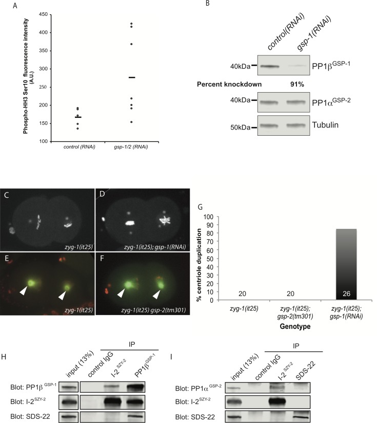
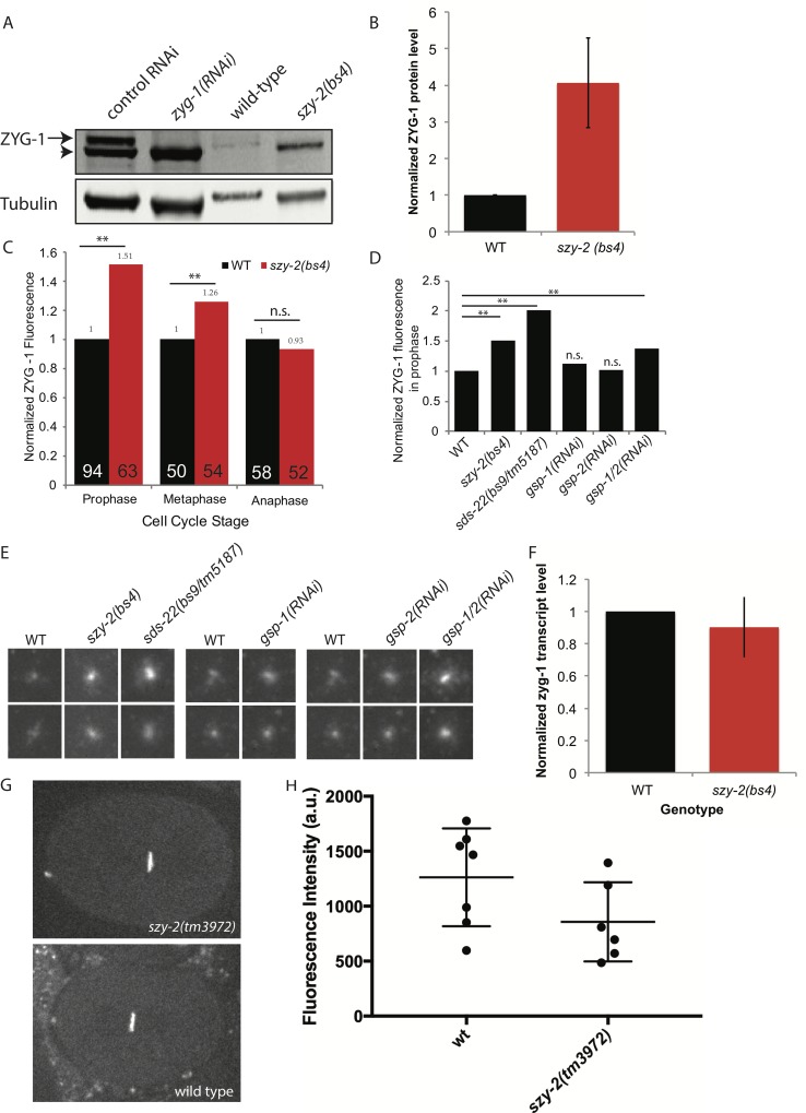
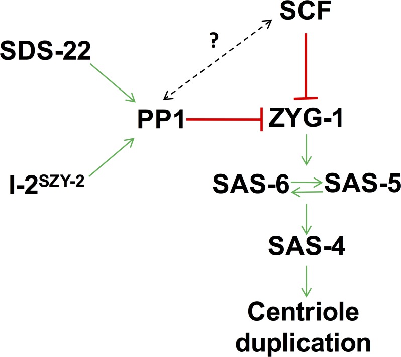
In humans perturbations of centriole number are associated with tumorigenesis and microcephaly, therefore appropriate regulation of centriole duplication is critical. The C. elegans homolog of Plk4, ZYG-1, is required for centriole duplication, but our understanding of how ZYG-1 levels are regulated remains incomplete. We have identified the two PP1 orthologs, GSP-1 and GSP-2, and their regulators I-2SZY-2 and SDS-22 as key regulators of ZYG-1 protein levels. We find that down-regulation of PP1 activity either directly, or by mutation of szy-2 or sds-22 can rescue the loss of centriole duplication associated with a zyg-1 hypomorphic allele. Suppression is achieved through an increase in ZYG-1 levels, and our data indicate that PP1 normally regulates ZYG-1 through a post-translational mechanism. While moderate inhibition of PP1 activity can restore centriole duplication to a zyg-1 mutant, strong inhibition of PP1 in a wild-type background leads to centriole amplification via the production of more than one daughter centriole. Our results thus define a new pathway that limits the number of daughter centrioles produced each cycle.


Conflict of interest statement

The authors have declared that no competing interests exist.

Figures

Fig 1
Fig 1. Loss-of-function mutations in the PP1 regulator I-2SZY-2 rescues the zyg-1(it25) phenotype.
A) Scale diagram of the structure of the szy-2 gene, indicating the location of the tm3972 deletion and the szy-2(bs4) splice site mutation. B) Western blot of embryo extracts from wild-type and szy-2(bs4) mutants showed a reduced level of the SZY-2 protein. C) Stills from movies of embryos expressing mCherry::H2B and GFP::tubulin grown at 24°C. Top, wild type; Middle, zyg-1(it25) mutant at the 2-cell stage following centriole duplication failure; bottom, zyg-1(it25); szy-2(bs4) double mutants at 2 cell-stage showing a rescue of the centriole duplication. D) Quantification of centriole duplication failure at 24°C in zyg-1(it25) mutants and in zyg-1(it25) mutants in which szy-2 activity has been down-regulated by mutation or RNAi. Number of centriole duplication events analyzed is indicated. E) Western blot of embryo extracts, showing levels of phospho-histone H3 in wild-type and szy-2(bs4) embryos. F) Phospho-histone H3 staining of wild-type and szy-2(bs4) embryos at first metaphase. Left, merge: DNA blue, Microtubules red, Phospho-histone H3 green. Right, phospho-histone H3.
Fig 2
Fig 2. Reducing SDS-22 activity rescues the zyg-1(it25) phenotype.
A) Scale diagram of the structure of the sds-22 gene, indicating the location of the tm5187 deletion and the sds-22(bs9) substitution. B) Quantification of centriole duplication when zyg-1(it25) is rescued by the sds-22(bs9) mutation or sds-22(RNAi). Number of centriole duplication events analyzed is indicated. C) Complementation test showing bs9 and tm5187 are allelic. Hermaphrodites of the indicated genotype were shifted to 25°C at the L4 stage and embryonic lethality was determined over the next 24 hours. The szy-6(bs9) allele was marked with the closely linked dpy-10(e128) mutation.
Fig 3
Fig 3. PP1 functions with I-2SZY-2 and SDS-22 to regulate centriole duplication.
A) Comparison of phospho-HH3 levels in control and PP1βGSP-1 & PP1αGSP-2 co-depleted embryos as determined by quantitative immunofluorescence microscopy. The mean is indicated by the bar. B) Western blot demonstrating specificity of gsp-1(RNAi). C&D) Stills from time-lapse recordings of embryos of the indicated genotypes, expressing GFP::histone and GFP::SPD-2 grown at the normally restrictive temperature of 24°C. E&F) Stills from time-lapse recordings of embryos of the indicated genotypes, expressing mCherry::histone and GFP::tubulin grown at the normally restrictive temperature of 24°C; centrosomes are indicated by arrow heads. G) Quantitation of centriole duplication in indicated strains. Number of centriole duplication events scored is indicated. H) Western blots of immunoprecipitated material from whole worm extracts using control IgG, I-2SZY-2, or PP1βGSP-1 antibodies, and probed with I-2SZY-2, PP1βGSP-1, or SDS-22 antibodies as indicated. I) Western blots of immunoprecipitated material from whole worm extracts using control IgG, I-2SZY-2, or SDS-22 antibodies, and probed with I-2SZY-2 or PP1αGSP-2, or SDS-22 antibodies.
Fig 4
Fig 4. ZYG-1 protein levels are elevated in the szy-2(bs4) mutant.
A) Western blot of embryo extracts from wild-type and szy-2(bs4) embryos probed with a ZYG-1 antibody. The ZYG-1 band (arrow) is identified by its absence in zyg-1(RNAi) extracts. Non-specific proteins (arrowhead) are also bound by the ZYG-1 antibody and probably represent contaminants from the E. coli strain used for RNAi feeding experiments. B) Normalized ZYG-1 protein levels from two independent experiments. Error bars indicate standard deviation. C) Relative levels of centriole-localized ZYG-1 in wild-type and szy-2(bs4) one-cell embryos made by quantitative immunostaining. Levels at each stage are normalized to the wild-type control. Number of centrosomes analyzed indicated inside each bar. **p<0.01; n.s. no significant difference (Student’s t-test). D) Centriole-localized ZYG-1 protein levels for embryos of indicated genotypes at first mitotic prophase. Levels normalized to the wild-type control. Number of centrosomes analyzed indicated inside each bar. **p<0.01; n.s. no significant difference from WT (Student’s t-test). E) Representative images of centrosomal ZYG-1 levels in prophase. Centrosomes from 1-cell stage embryos of the indicated genotypes are grouped with a wild type sample from the same experiment. F) Quantification of zyg-1 transcript levels in embryos by qRT-PCR. Shown is the average of two independent experiments. Error bars indicate standard deviation. G) Representative images of wild-type and szy-2(tm3972) embryos expressing GFP::histone driven by the zyg-1 promoter and 3’-UTR. H) Quantitation of GFP intensities with mean and standard deviations indicated. No difference between strains was found (Student’s t-test p>0.1).
Fig 5
Fig 5. Decreasing PP1 activity leads to centriole overduplication.
A) Frames from a time-lapse recording of an sds-22(bs9/tm5187) trans-heterozygous embryo expressing GFP::SPD-2 and mCherry::histone. Supernumerary centrosomes appear at the 4-cell stage. B) Quantification of centrosome over-duplication in sds-22(bs9/tm5187) and szy-2(tm3972) embryos during the first and second rounds of centriole duplication. For sds-22(bs9/tm5187), n = 31/77 (first round/second round) and for szy-2(tm3972), n = 42/68 (first round/second round) C) Immunoblot showing extent of knockdown of PP1βGSP-1 and PP1αGSP-2 in gsp-1; gsp-2 double RNAi embryos. D) Selected frames from a time-lapse recording of a gsp-1(RNAi); gsp-2(RNAi) embryo expressing GFP::histone and mCherry::SPD-2. Arrowheads indicate extra centrosomes. E) Quantitation of the frequency of supernumerary centrioles in embryos depleted of the indicated PP1 genes. F) Structured illumination microscopy (SIM) of a late 2-cell stage sds-22(bs9/tm5187) trans-heterozygous embryo reveals the presence of excess centrioles. The low magnification image on the left shows the positions of the centrioles stained for the centriole marker SAS-4. Images on the right are enlargements of the indicated centrosomes. G) Quantification of SIM data to indicate the number of centrioles observed per centrosome comparing wild type and sds-22(bs9/tm5187) transheterozygous mutants.
Fig 6
Fig 6. Model of how I-2SZY-2, SDS-22 and PP1 might cooperate with the SCF to regulate centriole duplication.
Our work shows that PP1, I-2SZY-2 and SDS-22 down regulate ZYG-1 levels to constrain centriole duplication. We speculate that this is a new mechanism of regulation that operates independently of the previously documented SCF-mediated regulation. We cannot rule out, however, an interplay between the two pathways (dotted line).

Similar articles

Cited by

References

    1. Fu J, Hagan IM, Glover DM. The centrosome and its duplication cycle. Cold Spring Harb Perspect Biol. 2015;7: a015800 10.1101/cshperspect.a015800 - DOI - PMC - PubMed
    1. Habedanck R, Stierhof Y-D, Wilkinson CJ, Nigg EA. The Polo kinase Plk4 functions in centriole duplication. Nat Cell Biol. 2005;7: 1140–1146. 10.1038/ncb1320 - DOI - PubMed
    1. Strnad P, Leidel S, Vinogradova T, Euteneuer U, Khodjakov A, Gönczy P. Regulated HsSAS-6 levels ensure formation of a single procentriole per centriole during the centrosome duplication cycle. Dev Cell. 2007;13: 203–213. 10.1016/j.devcel.2007.07.004 - DOI - PMC - PubMed
    1. Arquint C, Sonnen KF, Stierhof Y-D, Nigg EA. Cell-cycle-regulated expression of STIL controls centriole number in human cells. J Cell Sci. 2012;125: 1342–1352. 10.1242/jcs.099887 - DOI - PubMed
    1. Peel N, Stevens NR, Basto R, Raff JW. Overexpressing centriole-replication proteins in vivo induces centriole overduplication and de novo formation. Curr Biol. 2007;17: 834–843. 10.1016/j.cub.2007.04.036 - DOI - PMC - PubMed

Publication types

MeSH terms

LinkOut - more resources