. 2017 Jan 19;17(1):22.
doi: 10.1186/s12866-017-0932-7.

ChMob2 binds to ChCbk1 and promotes virulence and conidiation of the fungal pathogen Colletotrichum higginsianum

Affiliations

ChMob2 binds to ChCbk1 and promotes virulence and conidiation of the fungal pathogen Colletotrichum higginsianum

Johannes Schmidpeter et al. BMC Microbiol. .

Abstract

Background: Mob family proteins are conserved between animals, plants and fungi and are essential for the activation of NDR kinases that control crucial cellular processes like cytokinesis, proliferation and morphology.

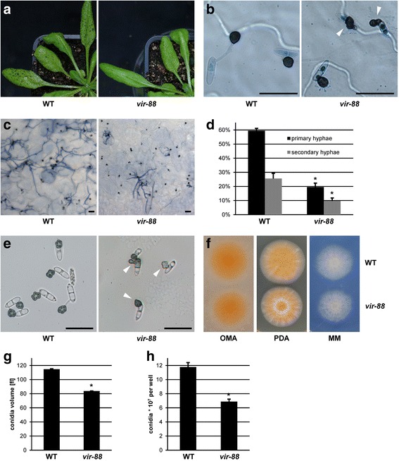
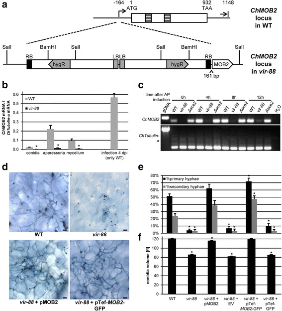
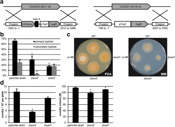
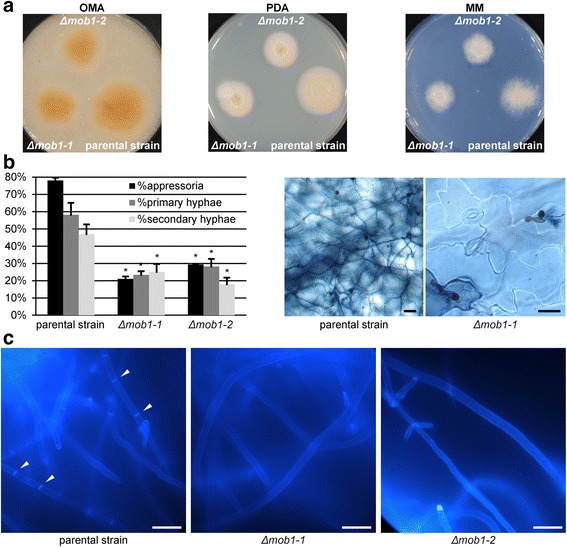
Results: We identified a hypomorphic allele of ChMOB2 in a random insertional mutant (vir-88) of the hemibiotrophic ascomycete fungus Colletotrichum higginsianum. The mutant is impaired in conidiation, host penetration and virulence on Arabidopsis thaliana. ChMob2 binds to and co-localizes with the NDR/LATS kinase homolog ChCbk1. Mutants in the two potential ChCbk1 downstream targets ChSSD1 and ChACE2 show defects in pathogenicity. The genome of C. higginsianum encodes two more Mob proteins. While we could not detect any effect on pathogenicity in ΔChmob3 mutants, ChMob1 is involved in conidiation, septae formation and virulence.

Conclusion: This study shows that ChMob2 binds to the conserved NDR/LATS Kinase ChCbk1 and plays an important role in pathogenicity of Colletotrichum higginsianum on Arabidopsis thaliana.

Keywords: ATMT; Ace2; Colletotrichum higginsianum; Conidiation; Cts1; Mob1; Mob2; Mob3; NDR Kinase Cbk1; Phytopathogenic ascomycete fungus; Ssd1.


Figures

Fig. 1
Fig. 1
T-DNA insertion mutant vir-88 is impaired in virulence and conidiation. A. thaliana Col-0 plants were infected with C. higginsianum WT and the vir-88 mutant strain. a Macroscopic image at 4 dpi. b Microscopic images of germinated conidia with appressoria at 1 dpi on trypan blue stained leaves. Appressoria with unusual morphology are marked with white arrows. c Trypan blue stained A. thaliana leaves at 3 dpi. d Quantification of trypan blue stained primary hyphae (relative to appressoria) and secondary hyphae (relative to primary hyphae) at 3 dpi. e Appressoria of WT and vir-88 on artificial hydrophobic surface. Appressoria with unusual morphology are marked with arrows. Scale bars = 20 μm. f Vegetative growth of WT and vir-88 on oatmeal agar (OMA), potato dextrose agar (PDA) and Czapek-Dox minimal medium (MM). g, h Average conidial cell volume g and conidial titer h of WT and vir-88 strains recovered from oatmeal agar after 7 days. The data of 3 separate plates is given. Error bars are represented as the standard error from three replicates. Significant differences based on t-tests (p < 0.05) are marked with asterisks
Fig. 2
Fig. 2
vir-88 encodes a hypomorphic ChMOB2 allele. a Schematic representation of the ChMOB2 locus in wild-type (top) and vir-88 (bottom). Beginning and end of the ChMOB2 transcript are shown relative to the start codon. Introns are depicted as striped boxes. LB = left border; RB = right border. b Quantitative RT-PCR of ChMOB2 mRNA abundance in conidia, appressoria, mycelium and in infected plants (4 dpi; in this case only data from WT is shown). The data were normalized against α-TUBULIN (CH063_01222) and are shown as the average of three biological replicates. c Semi-quantitative RT-PCR of ChMOB2 and α-TUBULIN in RNA samples from in vitro appressoria 0, 4, 8 and 12 h after inoculation. cDNAs from WT, vir-88 and ΔChace2 (CY6353, see below) strains were used as template for PCR. Genomic WT DNA (gDNA) or H2O were used as controls. d, e A. thaliana leaves at 3 days after infection with WT, vir-88 and vir-88 strains complemented with either the ChMOB2 wild-type allele (pMOB2, pCK3110) or with a pTef-MOB2-GFP construct (pCK4129). Typical trypan blue stained leaves are shown in d. Quantifications are shown in e. Scale bars = 20 μm. f Average volume of conidia from the indicated strains recovered from oatmeal agar after 7 days. The data from 3 separate plates is given. Error bars are represented as the standard error from three replicates. Significant differences based on t-tests (p < 0.05) are marked with asterisks
Fig. 3
Fig. 3
The genome of C. higginsianum encodes three Mob-like proteins. Unrooted phylogenetic tree of Mob proteins from C. higginsianum (marked in red) and other fungi. The tree was generated with the Geneious Tree builder plugin using a Blosum55 cost matrix and the Jukes-Cantor tree building method. The founding member of the Mob family (Phocein) from rat was included as a reference. Branch lengths correspond to substitutions per site. Scale bar = 0.2 substitutions per site
Fig. 4
Fig. 4
ChMob2 co-localizes with ChCbk1 in the cytoplasm. Conidia of ΔChcbk1:: CBK1-mCherry ΔChmob2:: MOB2-GFP (CY6720) were rinsed off oatmeal plates and analyzed by CLSM during in vitro appressoria formation. a Conidia, b in vitro AP at 8 hpi, c in vitro AP at 18 hpi. Bright field, GFP and mCherry channels are shown from left to right. The rightmost picture shows an overlay of GFP and mCherry channels. The intensities between panels are not directly comparable, because different settings were used to optimally visualize protein localization. Scale bars = 20 μm
Fig. 5
Fig. 5
ChMob2 interacts with ChCbk1. Whole cell extracts from strains expressing different combinations of ChMob2-GFP and ChCbk1-HA were used to analyze the interaction of ChMob2 and ChCbk1 by immunoprecipitation. Strains used from left to right: ΔChcbk1:: CBK1-HA pTef1a-MOB2-GFP (CY6681), the parental strain ΔChku80 (CY6021), ΔChcbk1:: CBK1-HA (CY6678) and pTef1a-MOB2-GFP (CY6543). Western blots of whole cell protein extracts (input), immunoprecipitates with anti-HA antibody coated beads (IP: α-HA) and immunoprecipitates with anti-GFP antibody coated beads (IP: α-GFP) were decorated either with anti-GFP (upper panels) or with anti-HA (lower panels) primary antibodies. The band corresponding to Mob2-GFP is marked with an arrow. The multiple bands corresponding to Cbk1-HA are marked with a bracket. Asterisks mark bands corresponding to primary antibody which are present in the IP and therefore are recognized by the secondary antibody
Fig. 6
Fig. 6
ChACE2 and ChSSD1 are required for full virulence of C. higginsianum. a Schematic representation of the genomic loci of ChACE2 and ChSSD1 (top) and the knockout cassettes of the respective deletion plasmids (bottom). The position of 5’ and 3’ flanking regions are given relative to the respective start codon. b Spray infection of A. thaliana at 3 dpi. Primary hyphae (relative to appressoria) and secondary hyphae (relative to primary hyphae) produced by the indicated strains. c Vegetative growth of the indicated strains on potato dextrose agar (PDA) and Czapek-Dox agar minimal medium (MM). d Average number of conidia (left) and cell volume (right) for the indicated strains recovered from oatmeal agar after 7 days. The data from 3 separate plates is shown. The strains used in b-d were wild-type (CY5535), vir-88, ΔChace2 (CY6353), ΔChssd1 (CY6649) and the ΔChku80 parental strain of the last two (CY6021). Error bars are represented as the standard error from three replicates. Significant differences based on t-tests (p < 0.05) are marked with asterisks
Fig 7
Fig 7
ChMOB1 is required for septum formation and cell division. Two independent ΔChmob1 transformants (CY7242 and CY7243) and the parental strain ΔChku80 (CY6021) were tested for vegetative growth, virulence and the ability to produce septae. a Vegetative growth on oatmeal agar (OMA), potato dextrose agar (PDA) and Czapek-Dox minimal medium (MM). b A. thaliana 4 days after droplet inoculation. Representative images of trypan blue stained leaves are shown on the right. The quantification of appressoria (relative to total conidia titer used for infection), primary hyphae (relative to appressoria) and secondary hyphae (relative to primary hyphae) of this infection is shown on the left. The data of 3 plants per strain with 2 leaves per plant with at least 100 counted appressoria per leaf is given. Error bars are represented as the standard error from three replicates. Significant differences based on t-tests (p < 0.05) are marked with asterisks. c Axenic mycelium stained with calcofluor white. Septae are marked with white arrows. Scale bars = 20 μm

Similar articles

Cited by

References

    1. Gupta YK, Dagdas YF, Martinez-Rocha AL, Kershaw MJ, Littlejohn GR, Ryder LS, Sklenar J, Menke F, Talbot NJ. Septin-Dependent Assembly of the Exocyst Is Essential for Plant Infection by Magnaporthe oryzae. Plant Cell. 2015;27:3277–89. doi: 10.1105/tpc.15.00552. - DOI - PMC - PubMed
    1. Giraldo MC, Dagdas YF, Gupta YK, Mentlak TA, Yi M, Martinez-Rocha AL, Saitoh H, Terauchi R, Talbot NJ, Valent B. Two distinct secretion systems facilitate tissue invasion by the rice blast fungus Magnaporthe oryzae. Nat Commun. 2013;4:1996. doi: 10.1038/ncomms2996. - DOI - PMC - PubMed
    1. Nesher I, Minz A, Kokkelink L, Tudzynski P, Sharon A. Regulation of pathogenic spore germination by CgRac1 in the fungal plant pathogen Colletotrichum gloeosporioides. Eukaryot Cell. 2011;10:1122–30. doi: 10.1128/EC.00321-10. - DOI - PMC - PubMed
    1. Sakaguchi A, Miyaji T, Tsuji G, Kubo Y. Kelch repeat protein Clakel2p and calcium signaling control appressorium development in Colletotrichum lagenarium. Eukaryot Cell. 2008;7:102–11. doi: 10.1128/EC.00227-07. - DOI - PMC - PubMed
    1. Fukada F, Kubo Y. Colletotrichum orbiculare Regulates Cell Cycle G1/S Progression via a Two-Component GAP and a GTPase to Establish Plant Infection. Plant Cell. 2015;27:2530–44. doi: 10.1105/tpc.15.00179. - DOI - PMC - PubMed

MeSH terms