

Stiffness of the locking compression plate as an external fixator for treating distal tibial fractures: a biomechanics study
- PMID: 28103852
- PMCID: PMC5248451
- DOI: 10.1186/s12891-016-1384-1
Stiffness of the locking compression plate as an external fixator for treating distal tibial fractures: a biomechanics study
Abstract
Background: Locking compress plate, as external fixator, is an attractive technique for distal tibial fracture treatment. But it still remains unclear whether the external LCP has sufficient stiffness. Thus, the present study aims to make a comprehensive evaluation of the stiffness of external locking compress plate when it is used as an external fixator in distal tibial fractures treatment.
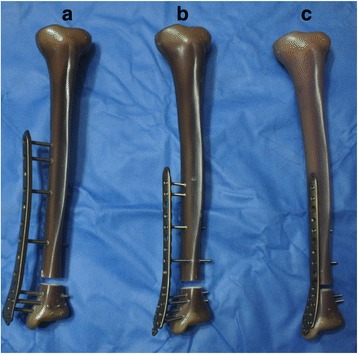
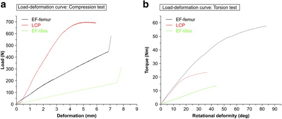
Methods: Composite tibia was used to simulate distal tibia fracture (Orthopedic Trauma Association type 43 A3 fracture). The fractures were stabilized with medial distal tibial locking compress plates (LCP group), medial distal tibial locking compress plates with 30-mm plate-bone distances (EF-tibia group), and medial distal femur locking compress plates with 30-mm plate-bone distances (EF-femur group). Stiffness of each configuration was measured under axial compression loading and in axial torsion loading directions. Compression stiffness and torsional rigidity were compared across different groups.
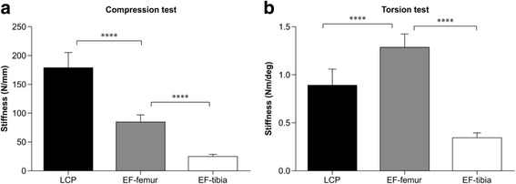
Results: Compared with LCP group, (1) EF-tibia group showed significantly lower (p < 0.001) compression stiffness and torsional rigidity; (2) EF-femur group showed significantly lower (p < 0.001) compression stiffness, but significantly higher (p < 0.001) torsional rigidity.
Conclusions: The results indicated that locking compress plate as an external fixator was flexible, and the distal femur locking compress plate was preferred over the distal tibial locking compress plate to be an external fixator in distal tibia fracture treatment.
Keywords: Biomechanics; External fixation; Fracture; Locking compression plate; Tibia.
Figures
Similar articles
-
Comparison between external locking plate fixation and conventional external fixation for extraarticular proximal tibial fractures: a finite element analysis.J Orthop Surg Res. 2022 Jan 11;17(1):16. doi: 10.1186/s13018-021-02907-3. J Orthop Surg Res. 2022. PMID: 35016716 Free PMC article.
-
Biomechanical study of the stiffness of the femoral locking compression plate of an external fixator for lower tibial fractures.BMC Musculoskelet Disord. 2023 Jan 18;24(1):39. doi: 10.1186/s12891-023-06150-1. BMC Musculoskelet Disord. 2023. PMID: 36650508 Free PMC article.
-
[A biomechanical study on internal and external fixation devices for treatment of humeral shaft fracture].Zhongguo Xiu Fu Chong Jian Wai Ke Za Zhi. 2008 May;22(5):516-9. Zhongguo Xiu Fu Chong Jian Wai Ke Za Zhi. 2008. PMID: 18630425 Chinese.
-
External fixation of the lower extremities: Biomechanical perspective and recent innovations.Injury. 2019 Jun;50 Suppl 1:S10-S17. doi: 10.1016/j.injury.2019.03.041. Epub 2019 Apr 5. Injury. 2019. PMID: 31018903 Review.
-
Hexapod External Fixation of Tibia Fractures in Children.J Pediatr Orthop. 2016 Jun;36 Suppl 1:S24-8. doi: 10.1097/BPO.0000000000000764. J Pediatr Orthop. 2016. PMID: 27078228 Review.
Cited by
-
Non‑contact locking plate: A useful alternative to external fixation in second‑stage treatment of post‑traumatic tibial osteomyelitis.Exp Ther Med. 2024 Mar 26;27(5):230. doi: 10.3892/etm.2024.12518. eCollection 2024 May. Exp Ther Med. 2024. PMID: 38596657 Free PMC article.
-
Comparison between external locking plate fixation and conventional external fixation for extraarticular proximal tibial fractures: a finite element analysis.J Orthop Surg Res. 2022 Jan 11;17(1):16. doi: 10.1186/s13018-021-02907-3. J Orthop Surg Res. 2022. PMID: 35016716 Free PMC article.
-
Investigating the biomechanical function of the plate-type external fixator in the treatment of tibial fractures: a biomechanical study.BMC Musculoskelet Disord. 2020 Feb 27;21(1):128. doi: 10.1186/s12891-020-3144-5. BMC Musculoskelet Disord. 2020. PMID: 32106851 Free PMC article.
-
A novel external fixation for treating tibial fractures: a finite element and biomechanical study.J Orthop Surg Res. 2025 Mar 28;20(1):319. doi: 10.1186/s13018-025-05681-8. J Orthop Surg Res. 2025. PMID: 40148896 Free PMC article.
-
A new classification and its value evaluation for intermediate column fractures of the distal radius.J Orthop Surg Res. 2018 Sep 3;13(1):221. doi: 10.1186/s13018-018-0925-8. J Orthop Surg Res. 2018. PMID: 30176895 Free PMC article.
References
-
- Nork SE, Schwartz AK, Agel J, Holt SK, Schrick JL, Winquist RA. Intramedullary nailing of distal metaphyseal tibial fractures. J Bone Joint Surg. 2005;87A(6):1213–21. - PubMed
Publication types
MeSH terms
LinkOut - more resources
Full Text Sources
Other Literature Sources
Medical
