. 2017 Jan 19;17(1):62.
doi: 10.1186/s12906-017-1583-9.

Sinularin induces DNA damage, G2/M phase arrest, and apoptosis in human hepatocellular carcinoma cells

Affiliations

Sinularin induces DNA damage, G2/M phase arrest, and apoptosis in human hepatocellular carcinoma cells

Ting-Wen Chung et al. BMC Complement Altern Med. .

Abstract

Background: Sinularin isolated from the cultured soft coral Sinularia flexibilis has been reported to exert potent cytotoxic effects against particular types of cancer. This study was carried out to investigate the cytotoxic effects in sinularin-treated human hepatocellular carcinoma cells, HepG2, and to subsequently explore the underlying molecular mechanisms.

Methods: TheMTT (3-[4,5-dimethylthiazol-2-yl]-2, 5-diphenyl- tetrazolium bromide) method was used to evaluate the cytotoxicity of sinularin on HepG2 and Hep3B cell lines. Furthermore, the cell cycle distribution assay, apoptosis assay, and western blot analysis in vitro were used to explore the possible mechanisms of action.

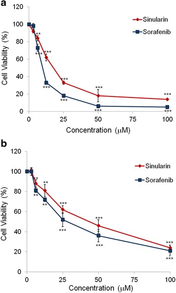
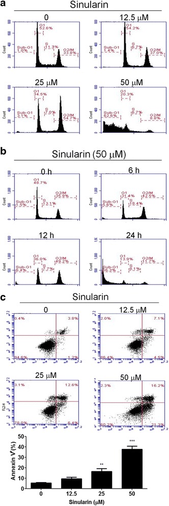
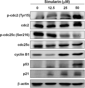
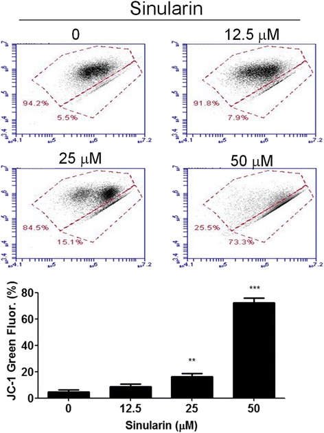
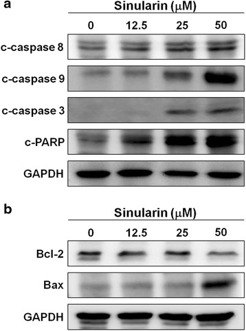
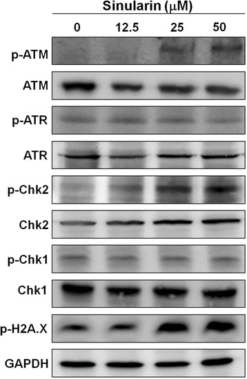
Results: From the results of our study, cell viability was obviously inhibited by sinularin in a dose-dependent manner. In addition, our results suggested that sinularin triggered DNA damage and subsequently induced cell cycle G2/M arrest associated with up-regulation of p-ATM (Ser(1981)), p-Chk2 (Tyr(68)), p-cdc2 (Tyr(15)), and p53 coupled with increased expression of downstream proteins p21 and down-regulation of p-cdc25 (Ser(216)). Moreover, the results of the apoptosis assay and western blot analysis indicated that the cytotoxic activity could be related to mitochondrial apoptosis, characterized by decrease of Bcl-2 expression, disruption of mitochondrial membrane potential, and sequential activation of caspases and Poly (ADP-ribose) polymerase (PARP).

Conclusions: This study reveals for the first time the anti-HCC activities of sinularin, the active compound isolated from the cultured soft coral Sinularia flexibilis. We believe that our results warrant further evaluation of sinularin as a new anti-HCC chemotherapeutic agent.

Keywords: Apoptosis; DNA damage; G2/M; Hepatocellular carcinoma; Sinularia flexibilis; Sinularin.


Figures

Fig. 1
Fig. 1
Analysis of cell viabilities in HCC cell lines. Percentages of cell viabilities of (a) HepG2 and (b) Hep3B cells after treating with sinularin or sorafenib at different concentrations for 24 h. Cell viabilities were plotted and represented as means ± SD. All data presented are representatives of three independent experiments with similar results. Significant differences from DMSO treated control group are indicated by **p < 0.01, ***p < 0.001
Fig. 2
Fig. 2
Cell cycle distributions of HepG2 cells with sinularin treatments. a Cell cycle analysis of sinularin-treated cells. HepG2 cells were treated with the indicated concentrations of sinularin for 24 h, or (b) treated with 50 μM of sinularin for 6, 12, and 24 h. Cell cycle distributions were determined by propidium iodide (PI) staining and flow cytometry analysis. Data are representative of three independent experiments with similar results. c Analysis of sinularin-induced apoptosis in HepG2 cells at indicated concentrations for 24 h. Phosphatidylserine externalization and DNA integrity were determined by FITC-annexin-V and PI, respectively. The lower-right quadrant (annexin-V+/PI) represents early apoptosis, while the upper-right quadrant (annexin V+/PI+) indicates late apoptosis and necrosis. The means ± SD of the experimental triplicates are presented in the bar graph at the bottom. All data presented are representatives of three independent experiments with similar results. Significant differences from DMSO treated control group are indicated by **p < 0.01, ***p < 0.001
Fig. 3
Fig. 3
Sinularin affected the cell cycle regulatory proteins in HePG2 cells. HepG2 cells were treated with different dosages of sinularin for 12 h and western blotting was performed to quantify the expression levels of cell cycle regulatory proteins. GAPDH expressions were measured as internal controls to show equal protein loading. The data presented are representatives of three independent experiments with similar results
Fig. 4
Fig. 4
Sinularin induced MMP reduction in HepG2 cells. HepG2 cells were treated with different dosages of sinularin for 24 h. Mitochondrial membrane potential (Δψm) was determined by JC-1 fluorescent dye staining and flow cytometry analysis. The means ± SD of the experimental triplicates were presented in the bar graph at the bottom. All data presented are representatives of three independent experiments with similar results. Significant differences from DMSO treated control group are indicated by **p < 0.01, ***p < 0.001
Fig. 5
Fig. 5
Sinularin regulated the protein levels of apoptosis in HepG2 cells. a and b Cell lysates of HepG2 cells were collected after sinularin treatments at 12.5, 25, and 50 μM for 24 h and western blotting was used to determine the protein expression levels. GAPDH expressions were measured as internal controls to show equal protein loading. The data presented are representatives of three independent experiments with similar results
Fig. 6
Fig. 6
Sinularin triggered DNA damage responses in HepG2 cells. HepG2 cells were treated with sinularin at 12.5, 25, and 50 μM for 24 h followed by western blotting to measure the DNA damage related proteins, phospho and non-phospho ATM, ATR, Chk1, Chk2, and H2A.X. GAPDH expressions were measured as internal controls to show equal protein loading. The data presented are representatives of three independent experiments with similar results

References

    1. Torre LA, Bray F, Siegel RL, Ferlay J, Lortet-Tieulent J, Jemal A. Global cancer statistics, 2012. CA Cancer J Clin. 2015;65:87–108. doi: 10.3322/caac.21262. - DOI - PubMed
    1. Altekruse SF, McGlynn KA, Reichman ME. Hepatocellular carcinoma incidence, mortality, and survival trends in the United States from 1975 to 2005. J Clin Oncol. 2009;27:1485–91. doi: 10.1200/JCO.2008.20.7753. - DOI - PMC - PubMed
    1. Moriguchi M, Umemura A, Itoh Y. Current status and future prospects of chemotherapy for advanced hepatocellular carcinoma. Clin J Gastroenterol. 2016;9:184–90. doi: 10.1007/s12328-016-0670-7. - DOI - PubMed
    1. Weinheimer AJ, Matson AJ, Hossain MB, van der Helm D. Marine anticancer agents: sinularin and dihydrosinularin, new cembranolides from the soft coral, sinularia flexibilis. Tetrahedron Lett. 1977;18:2923–6. doi: 10.1016/S0040-4039(01)83115-4. - DOI
    1. Su TR, Lin JJ, Chiu CC, Chen JY, Su JH, Cheng ZJ, et al. Proteomic investigation of anti-tumor activities exerted by sinularin against A2058 melanoma cells. Electrophoresis. 2012;33:1139–52. doi: 10.1002/elps.201100462. - DOI - PubMed

MeSH terms