. 2017 Jan 19;16(1):14.
doi: 10.1186/s12943-016-0570-y.

Aryl hydrocarbon receptor/cytochrome P450 1A1 pathway mediates breast cancer stem cells expansion through PTEN inhibition and β-Catenin and Akt activation

Affiliations

Aryl hydrocarbon receptor/cytochrome P450 1A1 pathway mediates breast cancer stem cells expansion through PTEN inhibition and β-Catenin and Akt activation

Abdullah Al-Dhfyan et al. Mol Cancer. .

Erratum in

Abstract

Background: Breast cancer stem cells (CSCs) are small sub-type of the whole cancer cells that drive tumor initiation, progression and metastasis. Recent studies have demonstrated a role for the aryl hydrocarbon receptor (AhR)/cytochrome P4501A1 pathway in CSCs expansion. However, the exact molecular mechanisms remain unclear.

Methods: The current study was designed to a) determine the effect of AhR activation and inhibition on breast CSCs development, maintenance, self-renewal, and chemoresistance at the in vitro and in vivo levels and b) explore the role of β-Catenin, PI3K/Akt, and PTEN signaling pathways. To test this hypothesis, CSC characteristics of five human breast cancer cells; SKBR-3, MCF-7, and MDA-MB231, HS587T, and T47D treated with AhR activators or inhibitor were determined using Aldefluor assay, side population, and mammosphere formation. The mRNA, protein expression, cellular content and localization of the target genes were determined by RT-PCR, Western blot analysis, and Immunofluorescence, respectively. At the in vivo level, female Balb/c mice were treated with AhR/CYP1A1 inducer and histopathology changes and Immunohistochemistry examination for target proteins were determined.

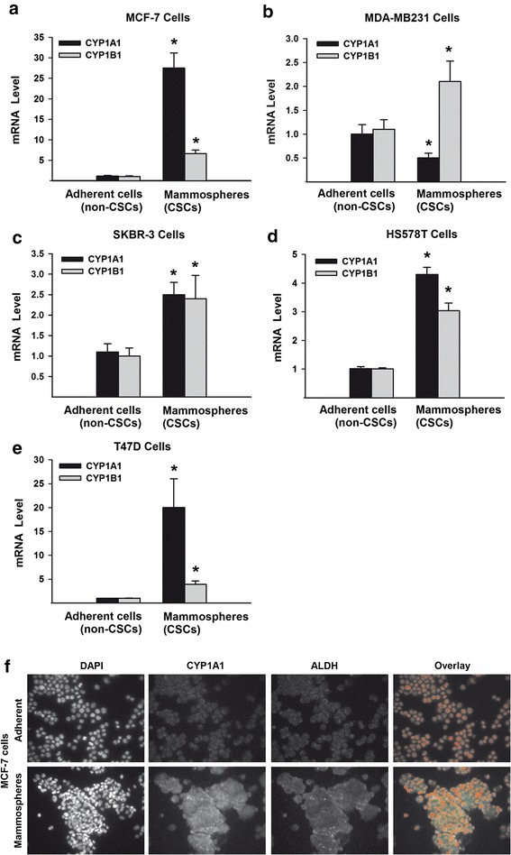
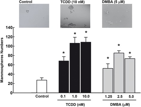
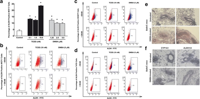
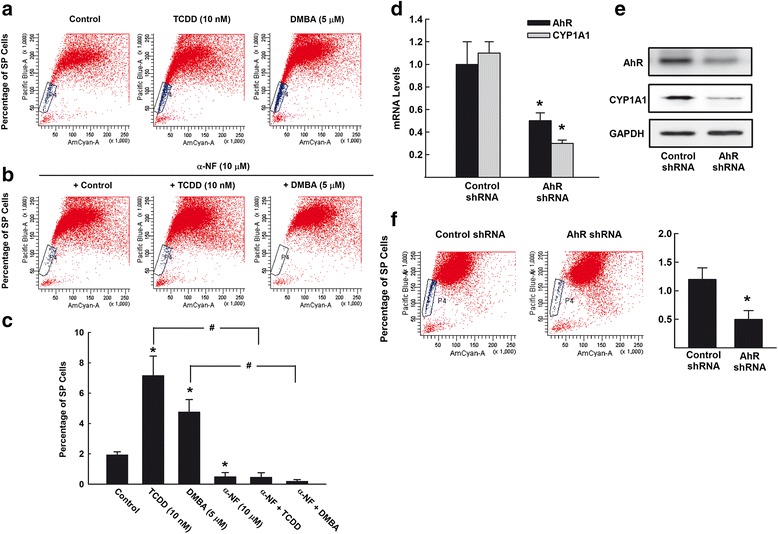
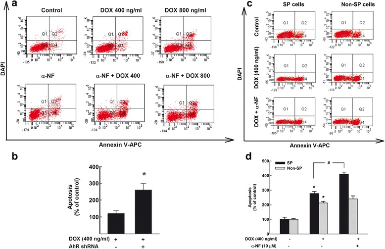
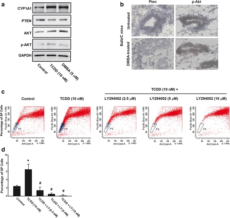
Results: The constitutive mRNA expression and cellular content of CYP1A1 and CYP1B1, AhR-regulated genes, were markedly higher in CSCs more than differentiating non-CSCs of five different human breast cancer cells. Activation of AhR/CYP1A1 in MCF-7 cells by TCDD and DMBA, strong AhR activators, significantly increased CSC-specific markers, mammosphere formation, aldehyde dehydrogenase (ALDH) activity, and percentage of side population (SP) cells, whereas inactivation of AhR/CYP1A1 using chemical inhibitor, α-naphthoflavone (α-NF), or by genetic shRNA knockdown, significantly inhibited the upregulation of ALDH activity and SP cells. Importantly, inactivation of the AhR/CYP1A1 significantly increased sensitization of CSCs to the chemotherapeutic agent doxorubicin. Mechanistically, Induction of AhR/CYP1A1 by TCDD and DMBA was associated with significant increase in β-Catenin mRNA and protein expression, nuclear translocation and its downstream target Cyclin D1, whereas AhR or CYP1A1 knockdown using shRNA dramatically inhibited β-Catenin cellular content and nuclear translocation. This was associated with significant inhibition of PTEN and induction of total and phosphorylated Akt protein expressions. Importantly, inhibition of PI3K/Akt pathway by LY294002 completely blocked the TCDD-induced SP cells expansion. In vivo, IHC staining of mammary gland structures of untreated and DMBA (30 mg/kg, IP)- treated mice, showed tremendous inhibition of PTEN expression accompanied with an increase in the expression p-Akt, β-Catenin and stem cells marker ALDH1.

Conclusions: The present study provides the first evidence that AhR/CYP1A1 signaling pathway is controlling breast CSCs proliferation, development, self-renewal and chemoresistance through inhibition of the PTEN and activation of β-Catenin and Akt pathways.

Keywords: AhR; Balb/c mice; Breast cancer; CYP1A1; Cancer stem cells; PI3K/Akt; PTEN; TCDD; shRNA; β-Catenin.


Figures

Fig. 1
Fig. 1
Constitutive expression of CYP1A1 and CYP1B1 in mammospheres vs adherent breast cancer cell lines. The basal mRNA expression of CYP1A1 and CYP1B1 of human breast cancer cell lines; MCF-7 (a), MDA-MB231 (b), SKBR-3 (c), HS578T (d), and T47D (e) were determined by RT-PCR normalized to β-ACTIN housekeeping gene. Duplicate reactions were performed for each experiment and the values are presented as mean ± SEM (n = 6). *; p < 0.05 compared to corresponding adherent cells. f Mammosphere and adherent populations of MCF-7 cells were stained with primary antibodies against CYP1A1 (green) and ALDH (magenta) followed by secondary antibodies and DAPI (red). Thereafter, the constitute CYP1A1 and ALDH proteins localization was conducted by Immunofluorescence assay. Each sample was stained in triplicate for each antibody
Fig. 2
Fig. 2
Effects of AhR activation on breast cancer mammospheres formation. MCF-7 cells were trated with indicated concentrations of TCDD and DMBA for 72 h. Thereafter, cells were trypsinized and viable 1000 cells/100μL were seeded into ultralow attachment plates in serum-free mammary epithelium basal medium, supplemented with B27, EGF, insulin, ABM, and hydrocortisone. Cells grown in these conditions as non-adherent spherical clusters (mammospheres) were allowed to grow for 7 days. The mammosphere cell numbers and size were determined by Evos® transmitted light microscope (Life Technologies Co., Grand Island, NY). Duplicate measurment were performed for each experiment and the values represent mean of fold change ± SEM (n = 3). *; p < 0.05 compared to control
Fig. 3
Fig. 3
Effects of AhR/CYP1A1 activation on breast CSCs marker ALDH in vitro and in vivo. a MCF-7 cells were treated for 72 h with increasing concentrations of TCDD (0.1, 1, 10 nM) and DMBA (1.25, 2.5, and 5 μM). b-d Three human breast cancer cells; MCF-7, HS587T, and T47D cells were treated for 72 h with TCDD 10 nM and DMBA 5 μM. Pelleted cells were then incubated with ALDH substrate in the presence and absence of DEAB, ALDH inhibitor. Thereafter, percentage of ALDH+ cells were determined by flow cytometry. Duplicate reactions were performed for each experiment. e-f Twenty virgin female Balb/c mice were injected IP with either corn oil (vehicle) or single dose of DMBA 30 mg/kg. Two months later, mammary gland tissues were collected and stained with H/E for histopathology (e) or with antibody against CYP1A1 and ALDH1/2 for IHC assay (f)
Fig. 4
Fig. 4
Effects of AhR/CYP1A1 inhibition on breast CSCs marker ALDH in vitro. a MCF-7 cells were treated for 72 h with TCDD 10 nM and DMBA 5 μM in the presence and absence of AhR/CYP1A1 inhibitor, α-NF 10 μM. Pelleted cells were then incubated with ALDH substrate in the presence and absence of DEAB, ALDH inhibitor. Thereafter, percentage of ALDH+ cells were determined by flow cytometry. b Duplicate reactions were performed for each experiment and the values represent mean of fold change ± SEM (n = 6). *; p < 0.05 compared to control. #; p < 0.05 compared to corresponding treatment in the absence of α-NF
Fig. 5
Fig. 5
Effects of AhR/CYP1A1 activation and inhibition on breast CSCs marker SP. a-c MCF-7 cells were treated for 72 h with TCDD 10 nM and DMBA 5 μM (a) or in the presence of α-NF 10 μM (b). Pelleted MCF-7 cells were incubated with DCV (10 μM) and the percentage of SP cells were then determined using LSRII® flow cytometer cell sorter. c The values represent mean ± SEM (n = 3). *; p < 0.05 compared to control. #; p < 0.05 compared to corresponding treatment in the absence of α-NF. d-f MCF-7 cells were stably transfected with specific AhR shRNA, thereafter, (d) the mRNA expression of AhR and CYP1A1 were quantified by RT-PCR normalized to β-ACTIN housekeeping gene. Duplicate reactions were performed for each experiment and the values are presented as mean ± SEM (n = 6). *p < 0.05 compared to corresponding control shRNA. e AhR and CYP1A1 protein expression levels were determined by Western blot analysis using the enhanced chemiluminescence method and one of three representative experiments is shown. f Pelleted MCF-7 cells were incubated with DCV (10 μM) and the percentage of SP cells were then determined using LSRII® flow cytometer cell sorter. The values represent mean ± SEM (n = 3). *; p < 0.05 compared to control shRNA
Fig. 6
Fig. 6
Effect of AhR inhibition on CSC apoptosis. a and b MCF-7 cells were treated for 72 h with DOX 400 or 800 ng/ml in the presence and absence of α-NF (10 μM) (a) or AhR shRNA (b). c and d SP and non-SP MCF-7 cells were treated for 72 h with DOX 400 ng/ml in the presence and absence of α-NF (10 μM). The cells were collected and then stained with annexin V–APC/DAPI. The percentage of cells underwent to apoptosis was then analysed on LSRII Flow Cytometer. One of the three representative experiments using different cell preparations was only shown. The values represent mean ± SEM (n = 3). *; p < 0.05 compared to control. #; p < 0.05 compared to DOX treatment
Fig. 7
Fig. 7
Effect of Wnt and Notch pathways inhibitors on TCDD-increased ALDH. a MCF-7 cells were treated for 72 h with TCDD 10 nM in the presence and absence of Wnt inhibitor (XAV-939, 5 μM) or Notch inhibitor (FLi 06, 5 μM). MCF-7 cell were then harvested, pelleted, and then incubated with ALDH substrate in the presence and absence of DEAB, ALDH inhibitor. Thereafter, percentage of ALDH+ cells were determined by flow cytometry. b MCF-7 cells treated with TCDD 10 nM were stained with primary antibodies against β-Catenin, Wnt transcription factor and ICN-1, Notch transcription factor followed by secondary antibodies. Thereafter, β-Catenin and ICN-1 proteins localization was conducted by immunofloursene assay. Each sample was stained in triplicate for each antibody
Fig. 8
Fig. 8
Effects of AhR/CYP1A1 activation and knockdown on β-Catenin levels in vitro and in vivo. a MCF-7 cells were treated with TCDD 10 nM and DMBA 5 μM for 72 h, thereafter the expression CYP1A1, β-Catenin, Cyclin D1 protein levels were determined by Western blot analysis using the enhanced chemiluminescence method and one of three representative experiments is shown. b MCF-7 cells treated with TCDD 10 nM were stained with primary antibodies against β-Catenin (green) followed by secondary antibodies and DAPI (red). Thereafter, β-Catenin localization and nuclear translocation was determined by immunofluorescence assay. Each sample was stained in triplicate for each antibody. c Twenty virgin female Balb/c mice were injected IP with either corn oil (vehicle) or single dose of DMBA 30 mg/kg. Two months later, mammary gland tissues were collected and stained with antibody against β-Catenin for IHC assay. d MCF-7 cells stably transfected with AhR shRNA (d) or CYP1A1 shRNA (e) were stained with primary antibodies against β-Catenin (green) and CYP1A1 (magenta) followed by secondary antibodies and DAPI (red). The β-Catenin and CYP1A1 localization and nuclear translocation were determined by immunofluorescence assay
Fig. 9
Fig. 9
Effect of AhR activation on PTEN/Akt pathway in vitro and in vivo. a MCF-7 cells were treated with TCDD 10 nM and DMBA 5 μM for 72 h, thereafter the expression CYP1A1, PTEN, Akt, and p-Akt protein levels were determined by Western blot analysis using the enhanced chemiluminescence method and one of three representative experiments is shown. b Twenty virgin female Balb/C mice were injected IP with either corn oil (vehicle) or single dose of DMBA 30 mg/kg. Two months later, mammary gland tissues were collected and stained with antibody against PTEN and p-Akt for determination by IHC assay. c and d MCF-7 cells were treated for 72 h with TCDD 10 nM in the presence and absence of increasing concentrations of Akt pathway inhibitor, LY294002. Thereafter, cells were harvested, pelleted, and then incubated with DCV (10 μM) and the percentage of SP cells were then determined using LSRII® flow cytometer cell sorter. The values represent mean ± SEM (n = 3). *; p < 0.05 compared to control, #; p < 0.05 compared to TCDD treatment

References

    1. Winquist RJ, Furey BF, Boucher DM. Cancer stem cells as the relevant biomass for drug discovery. Curr Opin Pharmacol. 2010;10:385–90. doi: 10.1016/j.coph.2010.06.008. - DOI - PubMed
    1. McDermott SP, Wicha MS. Targeting breast cancer stem cells. Mol Oncol. 2010;4:404–19. doi: 10.1016/j.molonc.2010.06.005. - DOI - PMC - PubMed
    1. Al-Hajj M, Wicha MS, Benito-Hernandez A, Morrison SJ, Clarke MF. Prospective identification of tumorigenic breast cancer cells. Proc Natl Acad Sci U S A. 2003;100:3983–8. doi: 10.1073/pnas.0530291100. - DOI - PMC - PubMed
    1. Bonnet D, Dick JE. Human acute myeloid leukemia is organized as a hierarchy that originates from a primitive hematopoietic cell. Nat Med. 1997;3:730–7. doi: 10.1038/nm0797-730. - DOI - PubMed
    1. Singh SK, Clarke ID, Terasaki M, Bonn VE, Hawkins C, Squire J, Dirks PB. Identification of a cancer stem cell in human brain tumors. Cancer Res. 2003;63:5821–8. - PubMed

Publication types

MeSH terms