

Manually defining regions of interest when quantifying paravertebral muscles fatty infiltration from axial magnetic resonance imaging: a proposed method for the lumbar spine with anatomical cross-reference
- PMID: 28103921
- PMCID: PMC5247810
- DOI: 10.1186/s12891-016-1378-z
Manually defining regions of interest when quantifying paravertebral muscles fatty infiltration from axial magnetic resonance imaging: a proposed method for the lumbar spine with anatomical cross-reference
Abstract
Background: There is increasing interest in paravertebral muscle composition as a potential prognostic and diagnostic element in lumbar spine health. As a consequence, it is becoming popular to use magnetic resonance imaging (MRI) to examine muscle volume and fatty infiltration in lumbar paravertebral muscles to assess both age-related change and their clinical relevance in low back pain (LBP). A variety of imaging methods exist for both measuring key variables (fat, muscle) and for defining regions of interest, making pooled comparisons between studies difficult and rendering post-production analysis of MRIs confusing. We therefore propose and define a method as an option for use as a standardized MRI procedure for measuring lumbar paravertebral muscle composition, and to stimulate discussion towards establishing consensus for the analysis of skeletal muscle composition amongst clinician researchers.
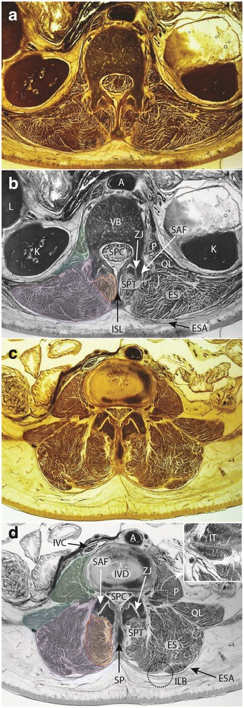
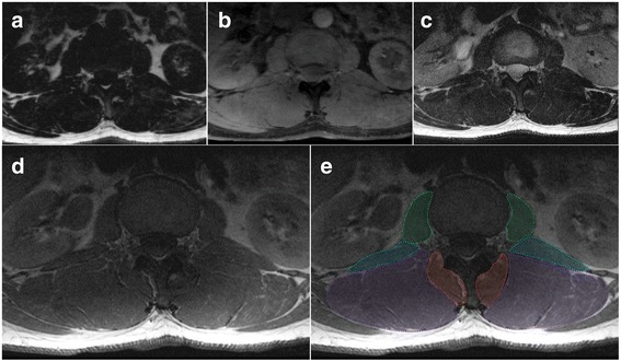
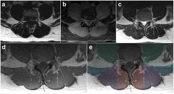
Method: In this descriptive methodological study we explain our method by providing an examination of regional lumbar morphology, followed by a detailed description of the proposed technique. Identification of paravertebral muscles and vertebral anatomy includes axial E12 sheet-plastinates from cadaveric material, combined with a series of axial MRIs that encompass sequencing commonly used for investigations of muscle quality (fat-water DIXON, T1-, and T2-weighted) to illustrate regional morphology; these images are shown for L1 and L4 levels to highlight differences in regional morphology. The method for defining regions of interest (ROI) for multifidus (MF), and erector spinae (ES) is then described.
Results: Our method for defining ROIs for lumbar paravertebral muscles on axial MRIs is outlined and discussed in relation to existing literature. The method provides a foundation for standardising the quantification of muscle quality that particularly centres on examining fatty infiltration and composition. We provide recommendations relating to imaging parameters that should additionally inform a priori decisions when planning studies examining lumbar muscle tissues with MRI.
Conclusions: We intend this method to provide a platform towards developing and delivering meaningful comparisons between MRI data on lumbar paravertebral muscle quality.
Keywords: Erector spinae; Fat infiltration; Lumbar spine; Magnetic resonance imaging; Manual segmentation; Multifidus; Paravertebral muscles; Region of interest.
Figures
Similar articles
-
Towards defining muscular regions of interest from axial magnetic resonance imaging with anatomical cross-reference: part II - cervical spine musculature.BMC Musculoskelet Disord. 2018 May 28;19(1):171. doi: 10.1186/s12891-018-2074-y. BMC Musculoskelet Disord. 2018. PMID: 29807530 Free PMC article.
-
Reliability of quantifying the spatial distribution of fatty infiltration in lumbar paravertebral muscles using a new segmentation method for T1-weighted MRI.BMC Musculoskelet Disord. 2016 May 27;17:234. doi: 10.1186/s12891-016-1090-z. BMC Musculoskelet Disord. 2016. PMID: 27230072 Free PMC article.
-
Methodological considerations in region of interest definitions for paraspinal muscles in axial MRIs of the lumbar spine.BMC Musculoskelet Disord. 2018 May 7;19(1):135. doi: 10.1186/s12891-018-2059-x. BMC Musculoskelet Disord. 2018. PMID: 29734942 Free PMC article.
-
Towards defining muscular regions of interest from axial magnetic resonance imaging with anatomical cross-reference: a scoping review of lateral hip musculature.BMC Musculoskelet Disord. 2022 Jun 4;23(1):533. doi: 10.1186/s12891-022-05439-x. BMC Musculoskelet Disord. 2022. PMID: 35658932 Free PMC article.
-
Fatty Infiltration in Paraspinal Muscles: Predicting the Outcome of Lumbar Surgery and Postoperative Complications.World Neurosurg. 2024 Oct;190:218-227. doi: 10.1016/j.wneu.2024.07.074. Epub 2024 Jul 15. World Neurosurg. 2024. PMID: 39019431 Review.
Cited by
-
The relation between local and distal muscle fat infiltration in chronic whiplash using magnetic resonance imaging.PLoS One. 2019 Dec 5;14(12):e0226037. doi: 10.1371/journal.pone.0226037. eCollection 2019. PLoS One. 2019. PMID: 31805136 Free PMC article.
-
The Effects of Reconditioning Exercises Following Prolonged Bed Rest on Lumbopelvic Muscle Volume and Accumulation of Paraspinal Muscle Fat.Front Physiol. 2022 Jun 14;13:862793. doi: 10.3389/fphys.2022.862793. eCollection 2022. Front Physiol. 2022. PMID: 35774286 Free PMC article.
-
Changes in Paraspinal Muscles and Facet Joints after Minimally Invasive Posterior Lumbar Interbody Fusion Using the Cortical Bone Trajectory Technique: A Prospective Study.Pain Res Manag. 2022 Jan 12;2022:2690291. doi: 10.1155/2022/2690291. eCollection 2022. Pain Res Manag. 2022. PMID: 35069954 Free PMC article.
-
The effect of high-intensity resistance exercise on lumbar musculature in patients with low back pain: a preliminary study.BMC Musculoskelet Disord. 2019 Jun 18;20(1):290. doi: 10.1186/s12891-019-2658-1. BMC Musculoskelet Disord. 2019. PMID: 31208400 Free PMC article. Clinical Trial.
-
Correlation of texture analysis of paraspinal musculature on MRI with different clinical endpoints: Lumbar Stenosis Outcome Study (LSOS).Eur Radiol. 2019 Jan;29(1):22-30. doi: 10.1007/s00330-018-5552-6. Epub 2018 Jun 14. Eur Radiol. 2019. PMID: 29948080 Clinical Trial.
References
-
- Brinjikji W, Luetmer PH, Comstock B, Bresnahan BW, Chen LE, Deyo RA, Halabi S, Turner JA, Avins AL, James K, et al. Systematic literature review of imaging features of spinal degeneration in asymptomatic populations. Am J Neuroradiol. 2015;36(4):811–816. doi: 10.3174/ajnr.A4173. - DOI - PMC - PubMed
-
- Amabile C, Moal B, Chtara OA, Pillet H, Raya JG, Iannessi A, Skalli W, Lafage V, Bronsard N. Estimation of spinopelvic muscles’ volumes in young asymptomatic subjects: a quantitative analysis. Surg Radiol Anat. 2016 Sep 16 [Epub ahead of print] doi:10.1007/s00276-016-1742-6. - PubMed
Publication types
MeSH terms
Grants and funding
LinkOut - more resources
Full Text Sources
Other Literature Sources
Medical
Research Materials
Miscellaneous
