. 2017 Jan 19;19(1):9.
doi: 10.1186/s13058-017-0801-1.

Elevated collagen-I augments tumor progressive signals, intravasation and metastasis of prolactin-induced estrogen receptor alpha positive mammary tumor cells

Affiliations

Elevated collagen-I augments tumor progressive signals, intravasation and metastasis of prolactin-induced estrogen receptor alpha positive mammary tumor cells

Craig E Barcus et al. Breast Cancer Res. .

Abstract

Background: The development and progression of estrogen receptor alpha positive (ERα+) breast cancer has been linked epidemiologically to prolactin. However, activation of the canonical mediator of prolactin, STAT5, is associated with more differentiated cancers and better prognoses. We have reported that density/stiffness of the extracellular matrix potently modulates the repertoire of prolactin signals in human ERα + breast cancer cells in vitro: stiff matrices shift the balance from the Janus kinase (JAK)2/STAT5 cascade toward pro-tumor progressive extracellular regulated kinase (ERK)1/2 signals, driving invasion. However, the consequences for behavior of ERα + cancers in vivo are not known.

Methods: In order to investigate the importance of matrix density/stiffness in progression of ERα + cancers, we examined tumor development and progression following orthotopic transplantation of two clonal green fluorescent protein (GFP) + ERα + tumor cell lines derived from prolactin-induced tumors to 8-week-old wild-type FVB/N (WT) or collagen-dense (col1a1 tm1Jae/+ ) female mice. The latter express a mutant non-cleavable allele of collagen 1a1 "knocked-in" to the col1a1 gene locus, permitting COL1A1 accumulation. We evaluated the effect of the collagen environment on tumor progression by examining circulating tumor cells and lung metastases, activated signaling pathways by immunohistochemistry analysis and immunoblotting, and collagen structure by second harmonic generation microscopy.

Results: ERα + primary tumors did not differ in growth rate, histologic type, ERα, or prolactin receptor (PRLR) expression between col1a1 tm1Jae/+ and WT recipients. However, the col1a1 tm1Jae/+ environment significantly increased circulating tumor cells and the number and size of lung metastases at end stage. Tumors in col1a1 tm1Jae/+ recipients displayed reduced STAT5 activation, and higher phosphorylation of ERK1/2 and AKT. Moreover, intratumoral collagen fibers in col1a1 tm1Jae/+ recipients were aligned with tumor projections into the adjacent fat pad, perpendicular to the bulk of the tumor, in contrast to the collagen fibers wrapped around the more uniformly expansive tumors in WT recipients.

Conclusions: A collagen-dense extracellular matrix can potently interact with hormonal signals to drive metastasis of ERα + breast cancers.

Keywords: Breast cancer; Collagen; Desmoplasia; Extracellular matrix; Prolactin; Tumor microenvironment; Tumor progression.


Figures
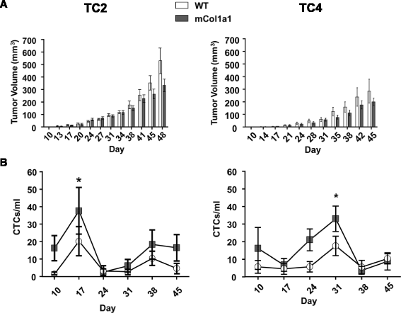
Fig. 1
Fig. 1
mCol1a1 does not alter the rate of tumor growth, but increases circulating tumor cells. a-b Tumor growth of tumor cell line (TC)2 (a) and TC4 (b) tumors until collection of the largest tumors began. Tumor volume was measured biweekly. Mean ± SEM TC2 animals: wild-type (WT) n = 7, mCol1a1 n = 6. TC4 animals: WT n = 7, mCol1a1 n = 8. Time significantly influenced tumor volume in both TC2 (a) and TC4 (b) tumors (p < 0.0001, two-way repeated measures ANOVA), but genotype did not. c-d Circulating tumor cells (CTCs) in peripheral blood of animals bearing TC2 (c) or TC4 (d) tumors. Peripheral blood was collected weekly and analyzed for green fluorescent protein positive (GFP+) CTCs as described in “Methods”. Mean ± SEM TC2 recipients: WT n = 7, mCol1a1 n =6. TC4 recipients: WT n = 7, mCol1a1 n =8. Genotype significantly altered levels of CTCs (TC2, p < 0.0050; TC4, p < 0.0320; repeated measure two-way ANOVA, taking into account values across days in the same animals). Levels of CTCs peaked when tumors were relatively small, regardless of genotype (TC2, day 17 is significantly different from all other days except day 38, *p < 0.05; TC4, day 31 is significantly higher than day 17 and day 38, *p < 0.05)
Fig. 2
Fig. 2
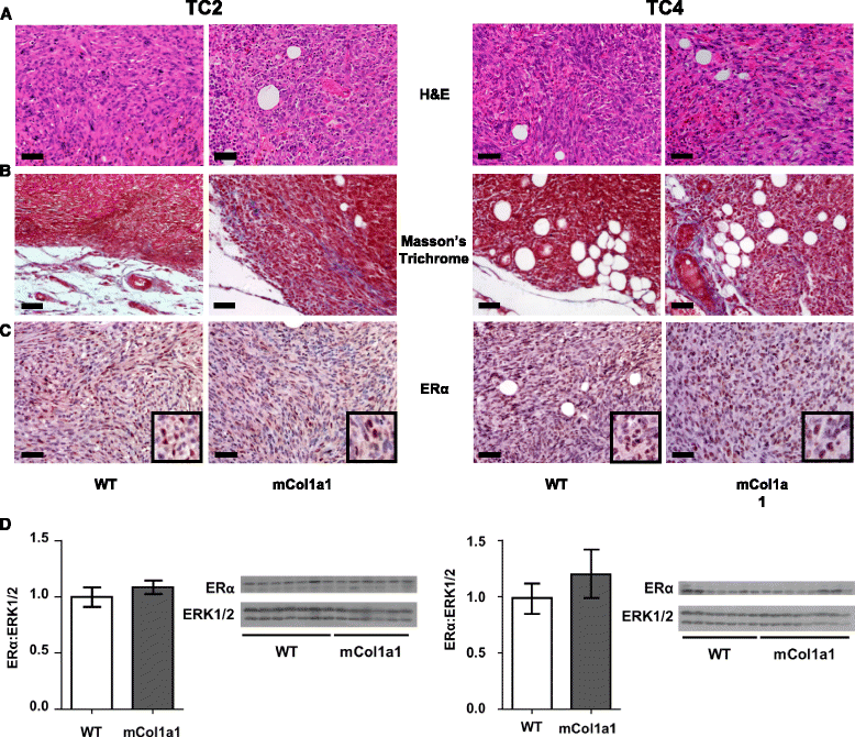
mCol1a1 does not alter tumor histologic type or hormone receptor status. Tumor cell line (TC)2 tumors (left panel) and TC4 tumors (right panel) were collected at end stage and processed for histological analysis as described in “Methods”. a Hematoxylin and eosin stain (H&E). b Masson’s Trichrome stain. c Estrogen receptor (ER)α immunohistochemical analysis. Original magnifications × 200; scale bar = 50 μm. d Immunoblotting of tumor lysates with the indicated antibodies. ERα levels were normalized to total extracellular regulated kinase (ERK)1/2. Note that increased intratumoral collagen-I in the mCol1a1 environment contributed to the protein harvested, reducing levels of signaling proteins when equal amounts of protein were loaded. Mean ± SEM TC2 tumors: wild-type (WT) n = 7, mCol1a1 n = 6; TC4 tumors: WT n = 7, mCol1a1 n = 8. Unpaired t test, p > 0.05
Fig. 3
Fig. 3
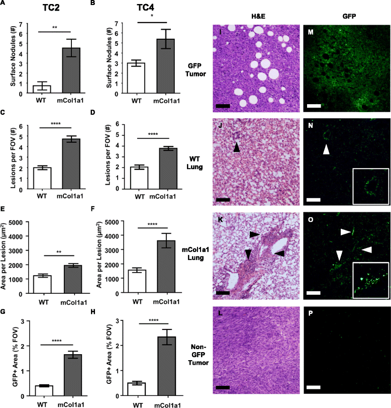
Metastasis of estrogen receptor (ER)α + tumors is enhanced by mCol1a1. a, b Nodules were counted on the surfaces of all lung lobes under dissection microscopy. Mean ± SEM tumor cell line (TC)2 tumors (a): wild-type (WT) n = 7, mCol1a1 n = 6. TC4 tumors (b): WT n = 7, mCol1a1 n = 8. Mann-Whitney U test, *p < 0.05, **p < 0.01. c-h Lungs were fixed and step-sectioned for histochemical analysis as detailed in “Methods”. Green fluorescent protein positive (GFP+) lesions were counted per × 10 field of view (FOV) (c, d). The area of GFP+ lesions was determined in ImageJ, and average size of individual lesions (e, f) and total lesion area per FOV (g, h) were calculated as described in “Methods”. Mean ± SEM Mann-Whitney U test. **p < 0.01, ****p < 0.0001. i-o Paired hematoxylin/eosin (H&E) and GFP stained sections from animals bearing TC4 tumors. Primary tumor (i, m), WT lung (j, n), mCol1a1 lung (k, o), and a non-GFP tumor control (l, p). Arrowheads indicate GFP+ lesions in lungs, one enlarged in each inset. Original magnifications × 100. Scale bar = 100 μm
Fig. 4
Fig. 4
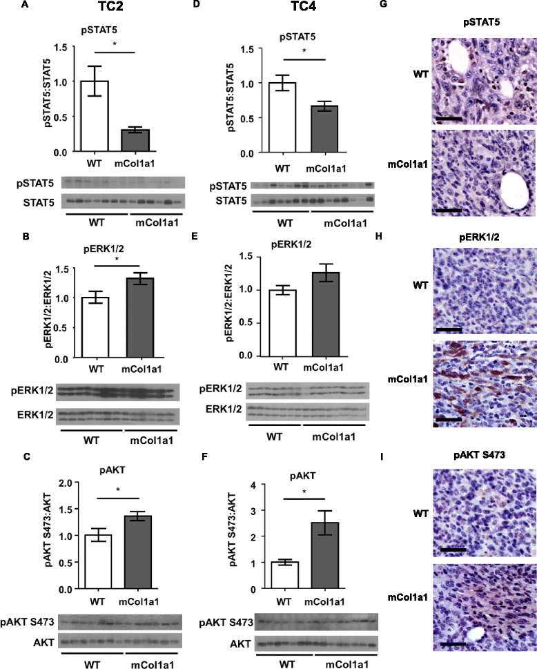
mCol1a1 decreases pSTAT5, and increases pERK1/2, pAKT. a-f TC2 (a-c) and tumor cell line (TC)4 (d-f) tumor lysates were analyzed by western blotting with the indicated antibodies. Mean ± SEM TC2 tumors: wild-type (WT) n = 7, mCol1a1 n = 6; TC4 tumors: WT n = 7, mCol1a1 n = 8. Unpaired t test, *p < 0.05. g-i Immunohistochemical analysis of pSTAT5 (g), pERK1/2 (h) and pAKT (i) in TC2 tumors. Original magnification × 200. Scale bar = 50 μm
Fig. 5
Fig. 5
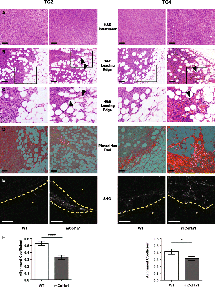
Early lesions in mCol1a1 recipients display invasive protrusions that contain aligned collagen fibers. a Hematoxylin/eosin stain (H&E) of tumor cell line (TC)2 tumors (left panels) at 17 days, TC4 tumors (right panels) at 24 days after transplantation. b Tumor edges (H&E). Arrowheads indicate finger-like protrusions into the mammary fat pad. c Higher magnification of boxed area from (b). d Picrosirius red staining. Note yellow-orange fibers in the protrusions in mCol1a1 tumors. Original magnification × 100. a, b, d, scale bars = 100 μm. e Second harmonic generation (SHG) imaging of collagen fibers. Yellow dashed lines indicate tumor boundaries. *Indicates adjacent fat pad. Representative images × 400 magnification. Scale bars = 50 μm. f CurveAlign analysis of relative collagen alignment following CT-FIRE fiber extraction as described in “Methods”. 0 = perfectly random fibers, 1 = perfectly aligned fibers. Mean ± SEM, n = 4 tumors of each cell line/host genotype combination, 7–9 images per tumor. Repeated measures, group comparison, SAS Proc Mixed model. *p < 0.05, ****p < 0.001. WT wild-type

References

    1. Anderson WF, Katki HA, Rosenberg PS. Incidence of breast cancer in the United States: current and future trends. J Natl Cancer Inst. 2011;103:1397–402. doi: 10.1093/jnci/djr257. - DOI - PMC - PubMed
    1. Voduc KD, Cheang MCU, Tyldesley S, Gelmon K, Nielsen TO, Kennecke H. Breast cancer subtypes and the risk of local and regional relapse. J Clin Oncol. 2010;28:1684–91. doi: 10.1200/JCO.2009.24.9284. - DOI - PubMed
    1. Early Breast Cancer Trialists’ Collaborative Group (EBCTCG) Effects of chemotherapy and hormonal therapy for early breast cancer on recurrence and 15-year survival: an overview of the randomised trials. Lancet. 2005;365:1687–717. doi: 10.1016/S0140-6736(05)66544-0. - DOI - PubMed
    1. Walker R. The complexities of breast cancer desmoplasia. Breast Cancer Res. 2001;3:143–5. doi: 10.1186/bcr287. - DOI - PMC - PubMed
    1. Butcher D, Alliston T, Weaver V. A tense situation: forcing tumour progression. Nat Rev Cancer. 2009;9:108–22. doi: 10.1038/nrc2544. - DOI - PMC - PubMed

Publication types

MeSH terms