Review
. 2017 Feb;33(2):86-100.
doi: 10.1016/j.tig.2016.12.004. Epub 2017 Jan 16.

Nuclear Dynamics of Heterochromatin Repair

Affiliations
Review

Nuclear Dynamics of Heterochromatin Repair

Nuno Amaral et al. Trends Genet. 2017 Feb.

Abstract

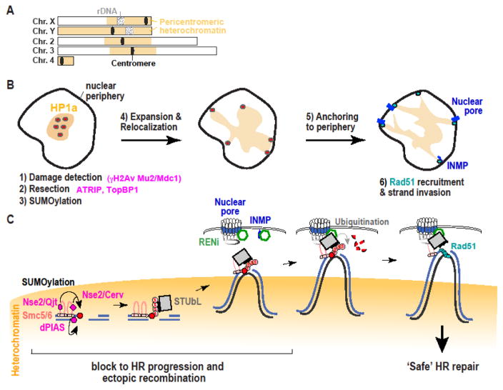
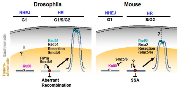
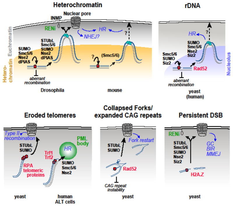
Repairing double-strand breaks (DSBs) is particularly challenging in pericentromeric heterochromatin, where the abundance of repeated sequences exacerbates the risk of ectopic recombination and chromosome rearrangements. Recent studies in Drosophila cells revealed that faithful homologous recombination (HR) repair of heterochromatic DSBs relies on the relocalization of DSBs to the nuclear periphery before Rad51 recruitment. We summarize here the exciting progress in understanding this pathway, including conserved responses in mammalian cells and surprising similarities with mechanisms in yeast that deal with DSBs in distinct sites that are difficult to repair, including other repeated sequences. We will also point out some of the most important open questions in the field and emerging evidence suggesting that deregulating these pathways might have dramatic consequences for human health.


Conflict of interest statement

The authors declare no competing financial interests.

Figures

Figure 1 (Key Figure)
Figure 1 (Key Figure). Model for the pathway that relocalizes heterochromatic DSBs to the nuclear periphery for continuing recombinational repair
A) Schematic view of Drosophila chromosomes showing the position and extension of pericentromeric heterochromatin (adapted from [29]). Cellular (B) and molecular (C) views show that when DSBs form in Drosophila heterochromatin (in yellow), early damage responses efficiently occur inside the domain. These likely include DSB detection, checkpoint activation, resection, and the recruitment of Smc5/6 and SUMO E3 ligases (i.e., Nse2/Qjt, Nse2/Cerv and dPIAS). SUMOylation of unknown targets blocks HR progression and ectopic recombination. SUMOylated proteins also recruit the STUbL protein Dgrn, and this might be sufficient to induce the relocalization to nuclear pores and INMPs at the nuclear periphery. The RENi protein dRad60 associates with STUbL at the nuclear periphery, promoting STUbL-mediated ubiquitination of SUMOylated targets, removal of the block to HR progression, Rad51 recruitment, and ‘safe’ HR repair. This removal of the block might rely on proteasome-mediated degradation of ubiquitinated target (as shown). Alternatively, these targets might become active after ubiquitination or de-SUMOylation (not shown). This model also predicts that sister chromatids or homologous chromosomes (black line) relocalize in concert with the damaged site to provide homologous templates for repair completion.
Figure 2
Figure 2. Comparison between Drosophila and mouse heterochromatin repair pathways
In both Drosophila and mouse cells, DSBs leave the heterochromatin domain during HR repair. Resection and the Smc5/6 complex are required for relocalization, and Rad51 is recruited after relocalization. Strand invasion components (including Rad51) mediate relocalization likely by stabilizing repair sites outside the heterochromatin domain. Mouse Smc5/6 also blocks NHEJ inside the domain. Heterochromatin expansion occurs during relocalization in both systems (not shown), potentially contributing to DSB signaling and repair and/or dynamics. In Drosophila cells, but not in mouse cells, Smc5/6 and HP1a are sufficient to block HR progression inside the heterochromatin domain, thus preventing ectopic recombination between heterochromatic sequences. Further, NHEJ is available as an alternative pathway for heterochromatin repair in G1; this pathway occurs without relocalization in mouse cells, but might require relocalization in Drosophila cells. Finally, Drosophila heterochromatic DSBs relocalize to the nuclear periphery to continue repair, while the final destination of this movement in mouse cells is still unclear. Whether STUbL and RENi proteins participate in heterochromatin repair in mouse cells is also unknown.
Figure 3
Figure 3. Overview of relocalization pathways and signaling mechanisms
The models show the molecular mechanisms responsible for relocalizing DSBs to sub-nuclear domains to continue repair, including the current understanding of the final destination of these movements, regulatory components of the SUMOylation pathway involved (black, bold), and repair outcomes (blue, italics). The potential role of SUMOylation/relocalization in genome stability is also shown (black, italics). Light blue circles indicate Rad51 recruitment and strand invasion. Question marks point to some of the questions highlighted in the main text. Whether SUMOylation contributes to the spatial and temporal regulation of rDNA repair in human cells is still unclear. GC: Gene conversion. BIR: Break-induced replication. MMEJ: Microhomology-mediated end joining.

References

    1. Peng JC, Karpen GH. Epigenetic regulation of heterochromatic DNA stability. Curr Opin Genet Dev. 2008;18(2):204–11. - PMC - PubMed
    1. Chiolo I, Tang J, Georgescu W, Costes SV. Nuclear dynamics of radiation-induced foci in euchromatin and heterochromatin. Mutat Res. 2013;750(1–2):56–66. - PMC - PubMed
    1. Hoskins RA, Carlson JW, Kennedy C, Acevedo D, Evans-Holm M, Frise E, Wan KH, Park S, Mendez-Lago M, Rossi F, Villasante A, Dimitri P, Karpen GH, Celniker SE. Sequence Finishing and Mapping of Drosophila melanogaster Heterochromatin. Science. 2007;316(5831):1625–1628. - PMC - PubMed
    1. Ho JW, Jung YL, Liu T, Alver BH, Lee S, Ikegami K, Sohn KA, Minoda A, Tolstorukov MY, Appert A, Parker SC, Gu T, Kundaje A, Riddle NC, Bishop E, Egelhofer TA, Hu SS, Alekseyenko AA, Rechtsteiner A, Asker D, Belsky JA, Bowman SK, Chen QB, Chen RA, Day DS, Dong Y, Dose AC, Duan X, Epstein CB, Ercan S, Feingold EA, Ferrari F, Garrigues JM, Gehlenborg N, Good PJ, Haseley P, He D, Herrmann M, Hoffman MM, Jeffers TE, Kharchenko PV, Kolasinska-Zwierz P, Kotwaliwale CV, Kumar N, Langley SA, Larschan EN, Latorre I, Libbrecht MW, Lin X, Park R, Pazin MJ, Pham HN, Plachetka A, Qin B, Schwartz YB, Shoresh N, Stempor P, Vielle A, Wang C, Whittle CM, Xue H, Kingston RE, Kim JH, Bernstein BE, Dernburg AF, Pirrotta V, Kuroda MI, Noble WS, Tullius TD, Kellis M, MacAlpine DM, Strome S, Elgin SC, Liu XS, Lieb JD, Ahringer J, Karpen GH, Park PJ. Comparative analysis of metazoan chromatin organization. Nature. 2014;512(7515):449–52. - PMC - PubMed
    1. Hoskins RA, Carlson JW, Wan KH, Park S, Mendez I, Galle SE, Booth BW, Pfeiffer BD, George RA, Svirskas R, Krzywinski M, Schein J, Accardo MC, Damia E, Messina G, Mendez-Lago M, de Pablos B, Demakova OV, Andreyeva EN, Boldyreva LV, Marra M, Carvalho AB, Dimitri P, Villasante A, Zhimulev IF, Rubin GM, Karpen GH, Celniker SE. The Release 6 reference sequence of the Drosophila melanogaster genome. Genome Res. 2015;25(3):445–58. - PMC - PubMed

Publication types