

Hepatic Prominin-1 expression is associated with biliary fibrosis
- PMID: 28104292
- PMCID: PMC5404955
- DOI: 10.1016/j.surg.2016.09.043
Hepatic Prominin-1 expression is associated with biliary fibrosis
Abstract
Background: Intrahepatic biliary fibrosis, as seen with cholestatic liver injuries such as biliary atresia, is mechanistically distinct from fibrosis caused by hepatocyte toxicity. We previously demonstrated the expansion of cells expressing the stem/progenitor cell marker Prominin-1, within regions of developing fibrosis in biliary atresia. Thus, we hypothesized that Prominin-1 expression is biliary fibrosis-specific.
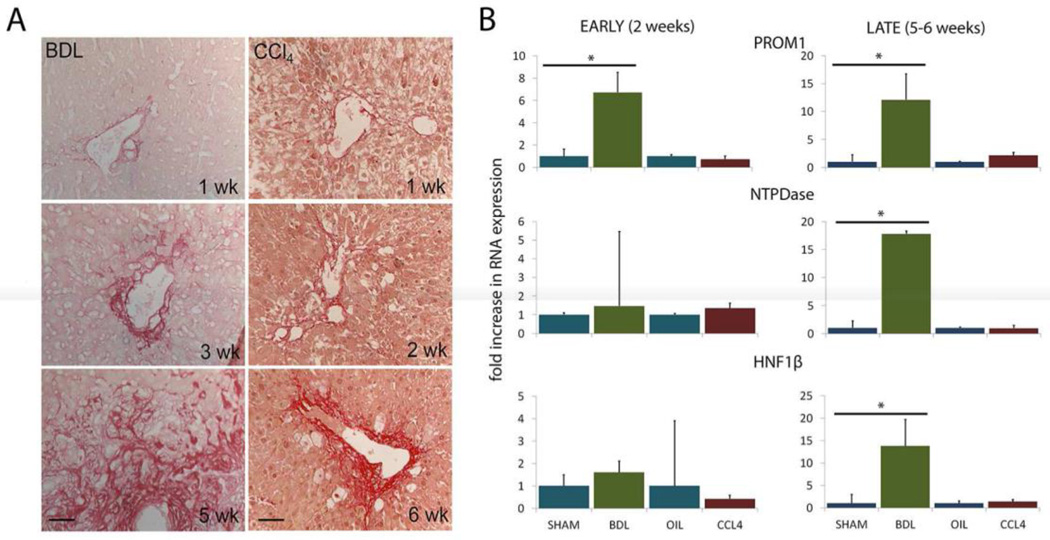
Methods: Gene expression of Prominin-1 was analyzed in adult mice undergoing either cholestatic bile duct ligation or hepatotoxic carbon tetrachloride administration by quantitative polymerase chair reaction. Lineage tracing of Prominin-1-expressing cells and Collagen-1α-expressing cells was performed after bile duct ligation in Prominin-1cre-ert2-lacz;Gfplsl and Collagen-1αGfp transgenic mice, respectively.
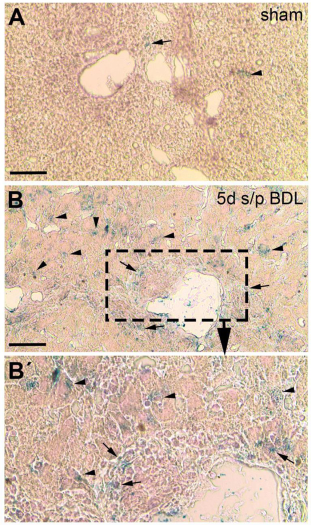
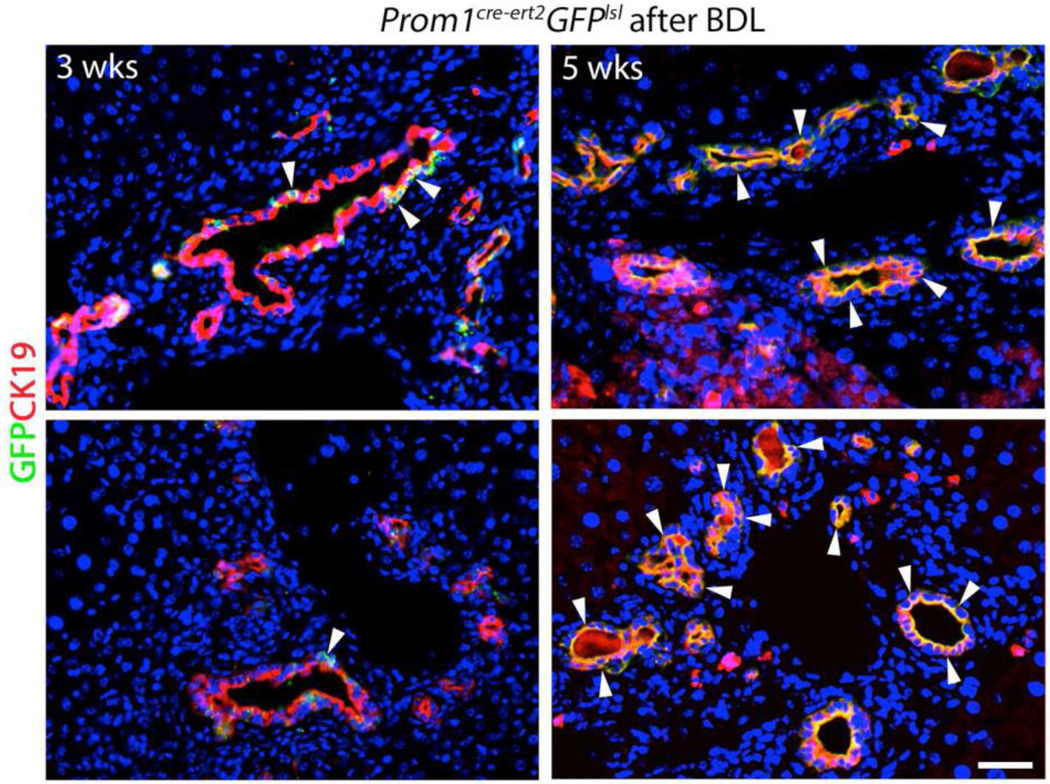
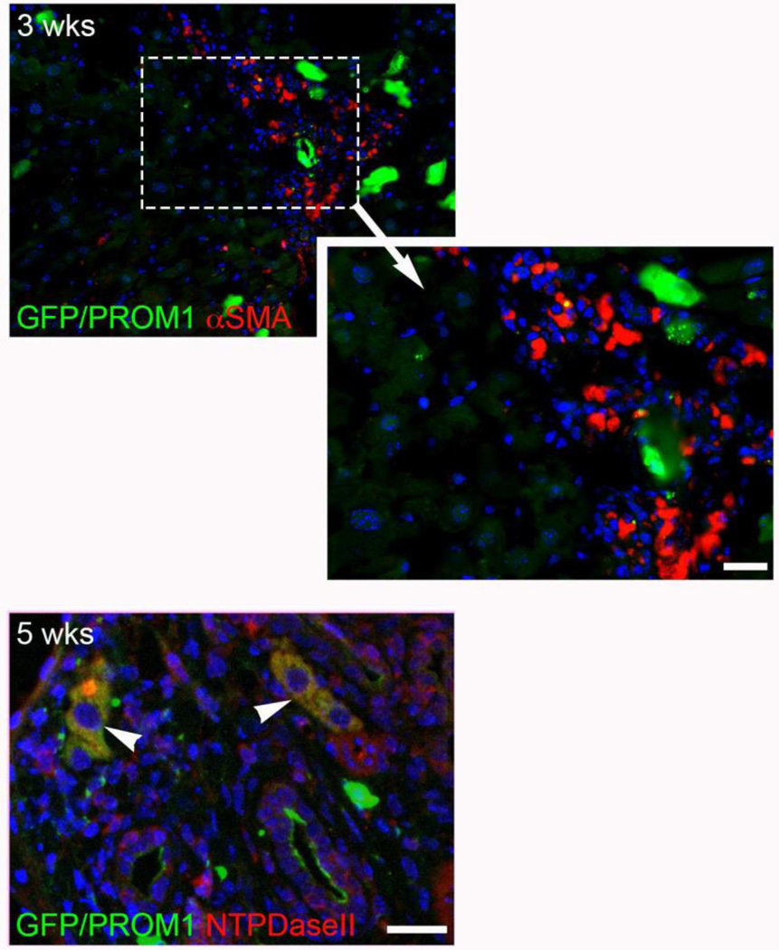
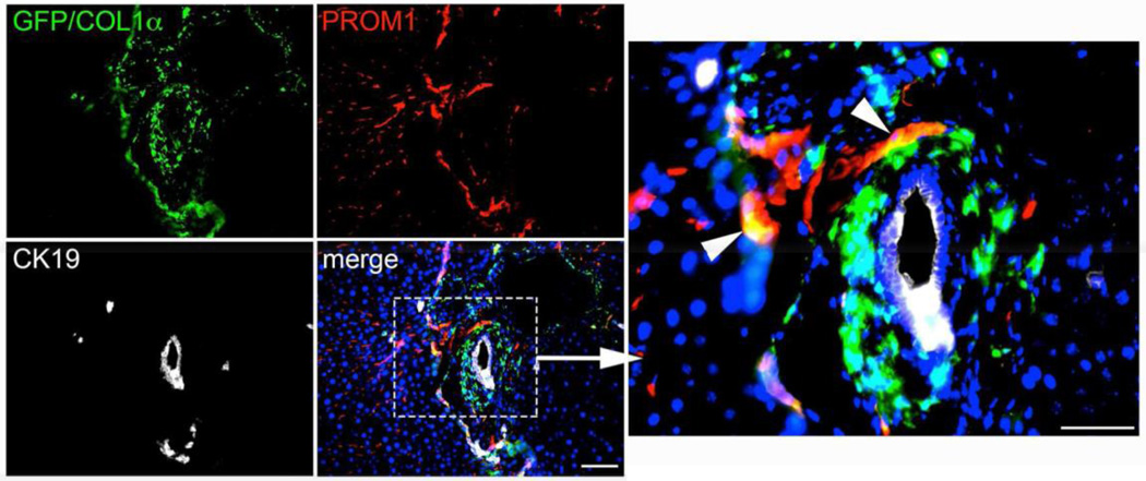
Results: Prominin-1 expression increased significantly after bile duct ligation compared with sham (6.6 ± 0.9-fold change at 2 weeks, P < .05) but not with carbon tetrachloride (-0.7 ± 0.5-fold change, not significant). Upregulation of Prominin-1 was observed histologically throughout the liver as early as 5 days after bile duct ligation in Prominin-1cre-ert2-lacz mice by LacZ staining in nonhepatocyte cells. Lineage tracing of Prominin-1-expressing cells labeled prior to bile duct ligation in Prominin-1cre-ert2-lacz;Gfplsl mice, demonstrated increasing colocalization of GREEN FLUORESCENT PROTEIN with biliary marker CYTOKERATIN-19 within ductular reactions up to 5 weeks after bile duct ligation consistent with biliary transdifferentiation. In contrast, rare colocalization of GREEN FLUORESCENT PROTEIN with mesenchymal marker α-SMOOTH MUSCLE ACTIN in Prominin-1cre-ert2-lacz;Gfplsl mice and some colocalization of GREEN FLUORESCENT PROTEIN with PROMININ-1 in Collagen-1αGfp mice, indicate minimal contribution of Prominin-1 progenitor cells to the pool of collagen-producing myofibroblasts.
Conclusion: During biliary fibrosis Prominin-1-expressing progenitor cells transdifferentiate into cells within ductular reactions. This transdifferentiation may promote fibrosis.
Copyright © 2016 Elsevier Inc. All rights reserved.
Figures
References
-
- Corbeil D, et al. The intriguing links between prominin-1 (CD133), cholesterol-based membrane microdomains, remodeling of apical plasma membrane protrusions, extracellular membrane particles, and (neuro)epithelial cell differentiation. FEBS Lett. 2010;584(9):1659–1664. - PubMed
-
- Marzesco AM, et al. Release of extracellular membrane particles carrying the stem cell marker prominin-1 (CD133) from neural progenitors and other epithelial cells. J Cell Sci. 2005;118(Pt 13):2849–2858. - PubMed
Publication types
MeSH terms
Substances
Grants and funding
LinkOut - more resources
Full Text Sources
Other Literature Sources
Research Materials
