. 2017 Apr 6;129(14):2013-2020.
doi: 10.1182/blood-2016-09-742999. Epub 2017 Jan 19.

Mice with a deficiency in CLEC-2 are protected against deep vein thrombosis

Affiliations

Mice with a deficiency in CLEC-2 are protected against deep vein thrombosis

Holly Payne et al. Blood. .

Abstract

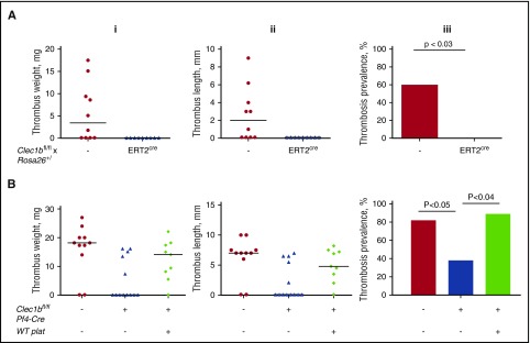
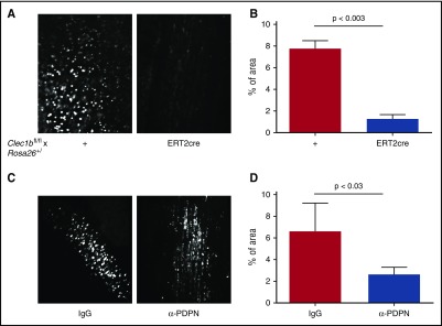
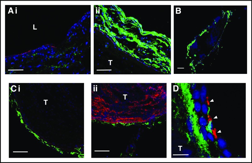
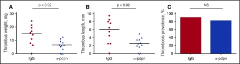
Deep vein thrombosis (DVT) with its major complication, pulmonary embolism, is a global health problem. Mechanisms of DVT remain incompletely understood. Platelets play a role in DVT, but the impact of specific platelet receptors remains unclear. Platelet C-type lectin-like receptor 2 (CLEC-2) is known to maintain the physiological state of blood vasculature under inflammatory conditions. DVT is a thromboinflammatory disorder developing largely as sterile inflammation in the vessel wall. We hypothesized therefore that CLEC-2 might play a role in DVT. Here, using a murine DVT model of inferior vena cava (IVC) stenosis, we demonstrate that mice with general inducible deletion of CLEC-2 or platelet-specific deficiency in CLEC-2 are protected against DVT. No phenotype in the complete stasis model was observed. Transfusion of wild-type platelets into platelet-specific CLEC-2 knockout mice restored thrombosis. Deficiency in CLEC-2 as well as inhibition of podoplanin, a ligand of CLEC-2, was associated with reduced platelet accumulation at the IVC wall after 6 hours of stenosis. Podoplanin was expressed in the IVC wall, where it was localized in the vicinity of the abluminal side of the endothelium. The level of podoplanin in the IVC increased after 48 hours of stenosis to a substantially higher extent in mice with a thrombus vs those without a thrombus. Treatment of animals with an anti-podoplanin neutralizing antibody resulted in development of smaller thrombi. Thus, we propose a novel mechanism of DVT, whereby CLEC-2 and upregulation of podoplanin expression in the venous wall trigger thrombus formation.


Figures

Figure 1.
Figure 1.
CLEC-2 exacerbates DVT in mice. (A) Clec1bfl/fl × Rosa26+/creERT2 (n = 10) and their Clec1bfl/fl littermates (n = 9) after 2 weeks of tamoxifen diet followed by 4 weeks of normal diet or (B) Clec1bfl/fl Pf4-Cre mice (n = 13) and control littermates (n = 11) were subjected to IVC stenosis for 48 hours. Some of the Clec1bfl/fl Pf4-Cre mice were transfused with 8 × 10⁸ WT platelets prior to surgery. (i-iii) Thrombus weight, thrombus length, and thrombosis prevalence, respectively. Lines in dot plots represent median. Note restoration of thrombosis after transfusion of WT platelets.
Figure 2.
Figure 2.
Stenosis-induced platelet recruitment is reduced in the absence of CLEC-2 or after neutralization of podoplanin. IVC stenosis was applied to Clec1bfl/fl × Rosa26+/creERT2 and their Clec1bfl/fl littermates for 6 hours. Fluorescently labeled syngeneic platelets were infused, and their deposition on the IVC wall was visualized by intravital microscopy. (A) Representative averaged images of adhered platelets (bright white). (B) Percentage of area covered by immobilized platelets, n = 3 to 4. Data are presented as mean ± standard error of the mean. (C) The same experimental design was applied to WT mice injected with anti–podoplanin neutralizing antibody (100 μg per mouse, IV) or IgG control before surgery. (D) Percentage of area covered by recruited platelets; n = 4 for both groups.
Figure 3.
Figure 3.
Podoplanin is expressed in the IVC vessel wall. Sham-operated IVC (Ai) or 48 stenosis-induced thrombi together with the IVC (Aii, B-C) were excised and stained for podoplanin (green) and PECAM-1 (Ai-ii, Ci; red) or VWF (Cii, red). Nuclei are blue in all images. (Ai-ii) Sham-operated IVC and IVC with a thrombus, respectively, photographed under identical microscope settings. Note increased podoplanin expression in the thrombosed IVC. (B) Whole thrombus after 48 hours of stenosis within the IVC. (Ci-ii) Costaining of podoplanin with PECAM-1 and VWF, respectively. Note podoplanin localization below the endothelium and absence of podoplanin in the thrombus. (D) Staining of IVC with a thrombus after 48 hours of stenosis for podoplanin (green) and platelets (CD41, red). Note platelets penetrating the vessel wall and localizing in the vicinity to podoplanin. Arrowheads indicate platelets. (A) Bar, 50 μm; (B) bar, 500 μm; (C) bar, 100 μm; n = 4-5. (D) Bar, 10 μm. Representative images of n = 3 with 10 to 15 images from each IVC. L, lumen; T, thrombus.
Figure 4.
Figure 4.
Expression of podoplanin in the IVC vessel wall increases with thrombosis. (A-C) Thrombi and the IVC were taken separately for western blotting. (A) Western blot of 5 thrombi vs 1 IVC after 48 hours of stenosis. Note complete absence of podoplanin in 4 of 5 thrombi and abundant podoplanin signal in the IVC. (B) Intact IVCs (lanes 1-3), IVCs after stenosis with thrombi (St+T, lanes 4-6), IVC after stenosis without thrombi (St N/T, lanes 7-9). Loading is confirmed by blotting for tubulin. (C) Densitometry of podoplanin bands normalized to tubulin; data in bar graph represent mean ± SD; n = 6 for each group. Ctrl, control.
Figure 5.
Figure 5.
Anti–podoplanin antibody decreases size of thrombi in murine DVT. WT mice were injected with neutralizing anti–podoplanin antibody (clone 8.1.1) 24 hours (100 μg per mouse) and 30 minutes (50 μg per mouse) prior to surgery and then subjected to IVC stenosis for 48 hours. Control mice were administered isotype-matched IgG. (A) Thrombus weight, (B) length, and (C) thrombosis prevalence. Lines in dot plots represent median. N = 10 (for IgG) and 11 (for the antibody). NS, not significant.

Comment in

References

    1. Astarita JL, Acton SE, Turley SJ. Podoplanin: emerging functions in development, the immune system, and cancer. Front Immunol. 2012;3:283. - PMC - PubMed
    1. Schacht V, Dadras SS, Johnson LA, Jackson DG, Hong YK, Detmar M. Up-regulation of the lymphatic marker podoplanin, a mucin-type transmembrane glycoprotein, in human squamous cell carcinomas and germ cell tumors. Am J Pathol. 2005;166(3):913-921. - PMC - PubMed
    1. May F, Hagedorn I, Pleines I, et al. . CLEC-2 is an essential platelet-activating receptor in hemostasis and thrombosis. Blood. 2009;114(16):3464-3472. - PubMed
    1. Suzuki-Inoue K, Inoue O, Ding G, et al. . Essential in vivo roles of the C-type lectin receptor CLEC-2: embryonic/neonatal lethality of CLEC-2-deficient mice by blood/lymphatic misconnections and impaired thrombus formation of CLEC-2-deficient platelets. J Biol Chem. 2010;285(32):24494-24507. - PMC - PubMed
    1. Hughes CE, Navarro-Núñez L, Finney BA, Mourão-Sá D, Pollitt AY, Watson SP. CLEC-2 is not required for platelet aggregation at arteriolar shear. J Thromb Haemost. 2010;8(10):2328-2332. - PMC - PubMed

Publication types