. 2017 Mar 14;135(11):1056-1070.
doi: 10.1161/CIRCULATIONAHA.116.024482. Epub 2017 Jan 19.

Long-Term Biased β-Arrestin Signaling Improves Cardiac Structure and Function in Dilated Cardiomyopathy

Affiliations

Long-Term Biased β-Arrestin Signaling Improves Cardiac Structure and Function in Dilated Cardiomyopathy

David M Ryba et al. Circulation. .

Abstract

Background: Biased agonism of the angiotensin II receptor is known to promote cardiac contractility. Our laboratory indicated that these effects may be attributable to changes at the level of the myofilaments. However, these signaling mechanisms remain unknown. Because a common finding in dilated cardiomyopathy is a reduction in the myofilament-Ca2+ response, we hypothesized that β-arrestin signaling would increase myofilament-Ca2+ responsiveness in a model of familial dilated cardiomyopathy and improve cardiac function and morphology.

Methods: We treated a dilated cardiomyopathy-linked mouse model expressing a mutant tropomyosin (Tm-E54K) for 3 months with either TRV120067, a β-arrestin 2-biased ligand of the angiotensin II receptor, or losartan, an angiotensin II receptor blocker. At the end of the treatment protocol, we assessed cardiac function using echocardiography, the myofilament-Ca2+ response of detergent-extracted fiber bundles, and used proteomic approaches to understand changes in posttranslational modifications of proteins that may explain functional changes. We also assessed signaling pathways altered in vivo and by using isolated myocytes.

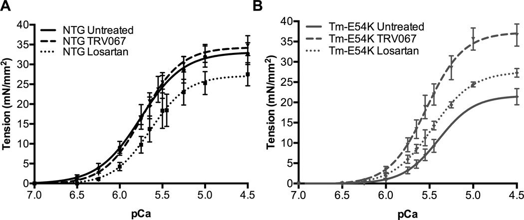
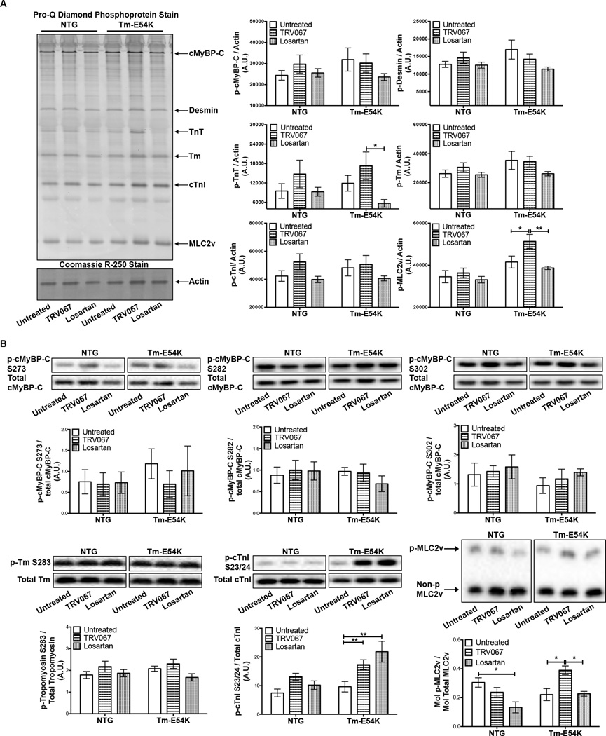
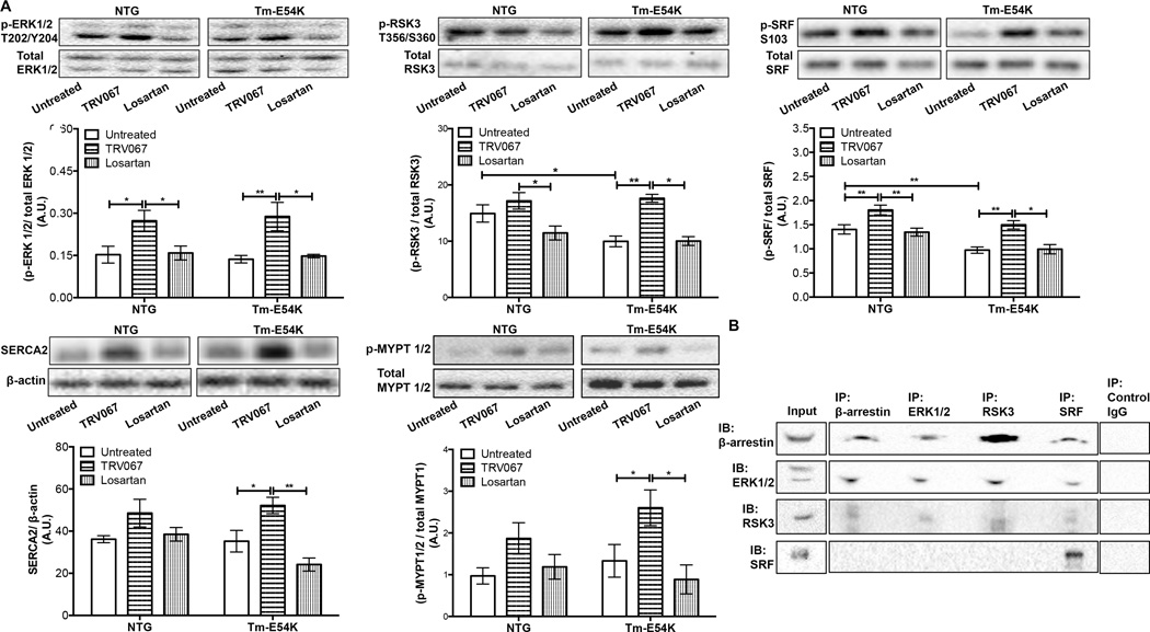
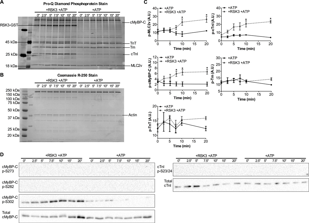
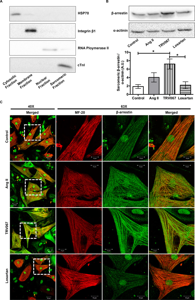
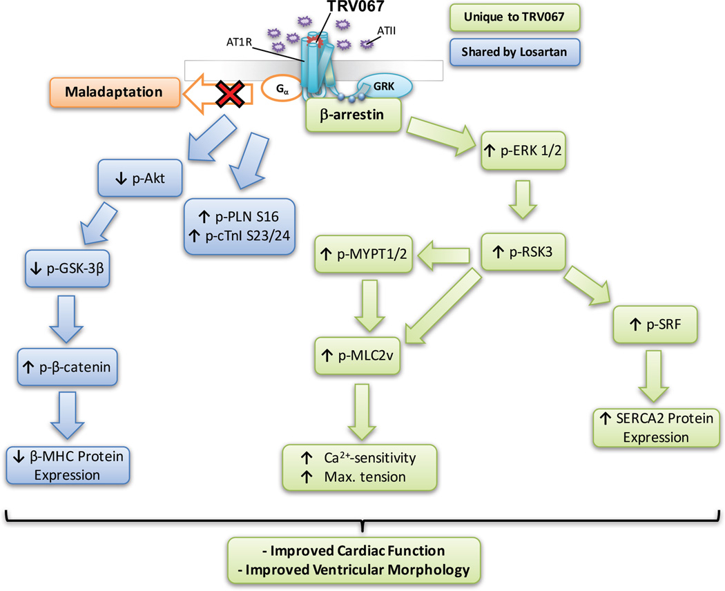
Results: TRV120067- treated Tm-E54K mice showed improved cardiac structure and function, whereas losartan-treated mice had no improvement. Myofilaments of TRV120067-treated Tm-E54K mice had significantly improved myofilament-Ca2+ responsiveness, which was depressed in untreated Tm-E54K mice. We attributed these changes to increased MLC2v and MYPT1/2 phosphorylation seen only in TRV120067-treated mice. We found that the functional changes were attributable to an activation of ERK1/2-RSK3 signaling, mediated through β-arrestin, which may have a novel role in increasing MLC2v phosphorylation through a previously unrecognized interaction of β-arrestin localized to the sarcomere.

Conclusions: Long-term β-arrestin 2-biased agonism of the angiotensin II receptor may be a viable approach to the treatment of dilated cardiomyopathy by not only preventing maladaptive signaling, but also improving cardiac function by altering the myofilament-Ca2+ response via β-arrestin signaling pathways.

Keywords: TRV120027; TRV120067; biased ligand; calcium sensitivity; sarcomeres.


Figures

Figure 1
Figure 1. TRV067 improved myofilament Ca2+-responsiveness and normalized tension generation in DCM
Force-Ca2+ relationship of fibers isolated from (A) NTG untreated, TRV067 and losartan treated animals and (B) Tm-E54K untreated, TRV067 and losartan treated animals following treatment for 3 months. Data were analyzed by two-way ANOVA followed by Tukey’s post-hoc test for multiple comparisons and represented as means ± SEM. N = 3–4 hearts per group, 6–8 fibers per group.
Figure 2
Figure 2. TRV067 and losartan induced post-translational modification of myofilament proteins
(A) Representative ProQ diamond and Coomassie R-250 stained gels and summarized analysis showing MLC2v phosphorylation was significantly increased in TRV067-treated Tm-E54Ks mice and decreased TnT phosphorylation in Tm-E54K losartan-treated mice. (B) Representative Western blots and summarized quantification of phospho-specific antibodies to cMyBP-C, Tm, and cTnI showing a significant increase in p-cTnI S23/24 in TRV067 and losartan-treated Tm-E54K mice and MLC2v phosphorylation significantly increased in TRV067-treated Tm-E54Ks mice and decreased in NTG losartan-treated mice. Data were analyzed by two-way ANOVA followed by Tukey’s post-hoc test for multiple comparisons and represented as means ± SEM. N = 4–6 hearts per group. * p ≤ 0.05. ** p ≤ 0.01
Figure 3
Figure 3. The Akt-GSK-3β pathway is downregulated in chronic TRV067 and losartan treated Tm-E54K mice
Representative Western blots and summarized quantification showing (A) Akt phosphorylation was decreased in TRV067 and losartan-treated Tm-E54K mice and GSK-3β phosphorylation was decreased. (B) There was a concomitant increase in β-catenin phosphorylation in these mice. (C) An increase in phosphorylation of phospholamban at S16 was also seen in both TRV067 and losartan-treated mice, but not at T17. Data were analyzed by two-way ANOVA followed by Tukey’s post-hoc test for multiple comparisons and represented as means ± SEM. N = 4–6 hearts per group. * p ≤ 0.05 , ** p ≤ 0.01, *** p ≤ 0.001.
Figure 4
Figure 4. TRV067, but not losartan, activated ERK1/2 and downstream signaling
Representative Western blot images and quantification showing (A) ERK1/2, RSK3, SRF phosphorylation, SERCA2 expression, and MYPT1/2 phosphorylation were all significantly increased in TRV067-treated mice but not losartan treated mice. (B) Representative western blot images of co-immunoprecipitation experiments of β-arrestin, ERK1/2, RSK3 and SRF. Data were analyzed by two-way ANOVA followed by Tukey’s post-hoc test for multiple comparisons and represented as means ± SEM. N = 4–6 hearts per group. * p ≤ 0.05 , ** p ≤ 0.01.
Figure 5
Figure 5. MLC2v, cTnI and cMyBP-C are substrates for RSK3 in vitro
Representative (A) Pro-Q diamond stained SDS-PAGE gel and (B) Coomassie R-250 stained SDS-PAGE gel showing an increase in MLC2v, cTnI, and cMyBPC by RSK3. (C) Summarized quantification of SDS-PAGE gels. (D) Western blot images showing phosphorylation of cMyBP-C at S302 by RSK3 but not S273 or S282 nor cTnI S23/24. Data were analyzed by repeated measures two-way ANOVA followed by Bonferroni’s post-hoc test for multiple comparisons and represented as means ± SEM. N = 4 hearts. * p ≤ 0.05.
Figure 6
Figure 6. A-band and M-line of the sarcomere show localization of β-arrestins, which was increased by TRV067
(A) Representative Western blots showing efficiency of subcellular fractionation in neonatal rat ventricular myocytes (NRVM). (B) Representative Western blot and summarized quantification of sarcomeric fractions of NRVMs that were treated for one hour with either angiotensin II (ang II), TRV067, or losartan and probed for β-arrestin and α-actinin. There is a basal amount of β-arrestin localized to the sarcomere under basal conditions, while analysis revealed that there was a significant increase β-arrestin localized to the sarcomere with TRV067 treatment. (C) Images of NRVMs stripped of their cytosolic and membrane components were stained for MF-20, light meromyosin, (red) or β-arrestin (green). Co-localization (yellow) which was increased by ang II and TRV067 treatment was observed for β-arrestin at the M-line and A-band, No increase was seen in the losartan -treated cells. Data were analyzed by one-way ANOVA followed by Tukey’s post-hoc test for multiple comparisons and represented as means ± SEM. N = 4 separate cultures. * p ≤ 0.05.
Figure 7
Figure 7. Proposed mechanism by which TRV067 improves cardiac function and morphology in DCM
Boxes in blue indicate pathways altered by both losartan and TRV067, whereas boxes in green indicate pathways only altered by TRV067.

Comment in

References

    1. Yancy CW, Jessup M, Bozkurt B, Butler J, Casey DE, Drazner MH, Fonarow GC, Geraci SA, Horwich T, Januzzi JL, Johnson MR, Kasper EK, Levy WC, Masoudi FA, McBride PE, McMurray JJV, Mitchell JE, Peterson PN, Riegel B, Sam F, Stevenson LW, Tang WHW, Tsai EJ, Wilkoff BL. 2013 ACCF/AHA Guideline for the Management of Heart Failure: A Report of the American College of Cardiology Foundation/American Heart Association Task Force on Practice Guidelines. Circulation. 2013;128:e240–e327. - PubMed
    1. Kim K-S, Abraham D, Williams B, Violin JD, Mao L, Rockman HA. β-Arrestin-biased AT1R stimulation promotes cell survival during acute cardiac injury. Am J Physiol Heart Circ Physiol. 2012;303:H1001–H1010. - PMC - PubMed
    1. Ahn S, Kim J, Hara MR, Ren XR, Lefkowitz RJ. β-Arrestin-2 Mediates Anti-apoptotic Signaling through Regulation of BAD Phosphorylation. J Biol Chem. 2009;284:8855–8865. - PMC - PubMed
    1. Correll CC, McKittrick BA. Biased Ligand Modulation of Seven Transmembrane Receptors (7TMRs): Functional Implications for Drug Discovery. J Med Chem. 2014;57:6887–6896. - PubMed
    1. Whalen EJ, Rajagopal S, Lefkowitz RJ. Therapeutic potential of beta-arrestin- and G protein-biased agonists. Trends Mol Med. 2011;17:126–139. - PMC - PubMed

MeSH terms