. 2017 Apr 11;135(15):1397-1412.
doi: 10.1161/CIRCULATIONAHA.116.023463. Epub 2017 Jan 19.

Detailed Echocardiographic Phenotyping in Breast Cancer Patients: Associations With Ejection Fraction Decline, Recovery, and Heart Failure Symptoms Over 3 Years of Follow-Up

Affiliations

Detailed Echocardiographic Phenotyping in Breast Cancer Patients: Associations With Ejection Fraction Decline, Recovery, and Heart Failure Symptoms Over 3 Years of Follow-Up

Hari K Narayan et al. Circulation. .

Abstract

Background: Cardiovascular disease in patients with breast cancer is of growing concern. The longitudinal effects of commonly used therapies, including doxorubicin and trastuzumab, on cardiac remodeling and function remain unknown in this population. We aimed to define the changes in echocardiographic parameters of structure, function, and ventricular-arterial coupling, and their associations with left ventricular ejection fraction (LVEF) and heart failure symptoms.

Methods: In a longitudinal prospective cohort study of 277 breast cancer participants receiving doxorubicin (Dox), trastuzumab (Tras), or both (Dox+Tras), we obtained 1249 echocardiograms over a median follow-up of 2.0 (interquartile range, 1.0-3.0) years. Left ventricular structure, diastolic and contractile function, and ventricular-arterial coupling measures were quantified in a core laboratory blinded to participant characteristics. We evaluated changes in echocardiographic parameters over time, and used repeated-measures regression models to define their association with LVEF decline and recovery. Linear regression models defined the association between early changes in these parameters and subsequent changes in LVEF and heart failure symptoms.

Results: Overall, 177 (64%) received Dox, 51 (18%) received Tras, and 49 (18%) received Dox+Tras. With Dox, there was a sustained, modest decrease in LVEF over the follow-up duration (1-year change in LVEF -3.6%; 95% confidence interval [CI], -4.4% to -2.8%; 3-year change -3.8%; 95% CI, -5.1% to -2.5%). With Tras, a similar LVEF decline was observed at 1 year (-4.5%; 95% CI, -6.0% to -2.9%) and 3 years (-2.8%; 95%CI, -5.3 to -0.4%). Participants receiving Dox+Tras demonstrated the greatest declines at 1 year (-6.6%; 95% CI, -8.2 to -5.0%), with partial recovery at 3 years (-2.8%; 95% CI, -4.8 to -0.8%). LVEF declines and recovery were associated primarily with changes in systolic volumes, longitudinal and circumferential strain, and ventricular-arterial coupling indices, effective arterial elastance (Ea) and the coupling ratio Ea/Eessb, without evidence for effect modification across therapies. Early changes in volumes, strain, and Ea/Eessb at 4 to 6 months were associated with 1- and 2-year LVEF changes. Similarly, early changes in strain and Ea were associated with worsening heart failure symptoms at 1 year.

Conclusions: Doxorubicin and trastuzumab resulted in modest, persistent declines in LVEF at 3 years. Changes in volumes, strain, and ventricular-arterial coupling were consistently associated with concurrent and subsequent LVEF declines and recovery across therapies.

Keywords: cardiotoxicity; doxorubicin; echocardiography; medical oncology; trastuzumab.


Figures

Figure 1
Figure 1. Changes in Left Ventricular Ejection Fraction by Treatment Regimen and After the Occurrence of Cardiac Dysfunction
(A) Changes in left ventricular ejection fraction (LVEF) are depicted over 3 years from the time of the baseline echocardiogram prior to cancer therapy initiation for each individual participant (grey). Loess smoothing splines representing changes in LVEF are displayed for participants receiving Doxorubicin (Dox, orange), Trastuzumab (Tras, green), and Doxorubicin+Trastuzumab (Dox+Tras, purple). (B) Changes in LVEF depicted for each individual participant after the occurrence of cardiac dysfunction plotted in grey. The dark black line represents the summary spline curve for LVEF across participants who experienced cardiac dysfunction.
Figure 2
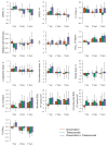
Figure 2. Changes in Left Ventricular Structure, Function, and Ventricular-Arterial Coupling by Treatment Regimen
Bars correspond to the marginal mean obtained from a repeated-measures linear regression model for the change in each echocardiographic parameter at 1, 2, and 3 years from baseline, by treatment regimen. Error bars represent 95% confidence intervals. Participants received Doxorubicin (Dox, orange), Trastuzumab (Tras, green), and Doxorubicin+Trastuzumab (Dox+Tras, purple). Measures include (A) LV end-diastolic volume (LVEDV); (B) LV end-systolic volume (LVESV); (C) LV mass; (D) relative wall thickness; (E) diastolic function (E/e′); (F) LV end-systolic elastance (Eessb); (G) longitudinal strain; (H) circumferential strain; (I) radial strain; (J) effective arterial elastance (Ea); (K) meridional end-systolic stress (ESS); (L) circumferential ESS; and (M) ventricular-arterial coupling ratio (Ea/Eessb).
Figure 3
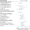
Figure 3. Associations between Echocardiographic Parameters and Changes in Left Ventricular Ejection Fraction According to Treatment Regimen
Point estimates and confidence intervals for the associations between changes in echocardiographic parameters and changes in left ventricular ejection fraction (LVEF) are graphically represented for participants receiving Doxorubicin (orange), Trastuzumab (green), and Doxorubicin+Trastuzumab (purple). Data also displayed in tabular format in the Supplemental File. Each point estimate corresponds to the absolute change in LVEF for each interquartile range increase in the echocardiographic parameter (shown in the leftmost column and estimated at the baseline visit), averaged across all followup timepoints. Interaction P values evaluate equality in associations across cancer therapy regimens. As an example, a point estimate of −1.5 corresponds to a 1.5% decrease in LVEF per interquartile range increase (27ml) in end-diastolic volume over the entire duration of followup. For longitudinal and circumferential strain, which are negative values, an increase represents a change from, for example, −15.7% to −11.8%. E/e′ = diastolic function index; Ea = effective arterial elastance; Eessb = end-systolic elastance; ESS = end-systolic stress; IQR = interquartile range; LV = left ventricle; LVEF = left ventricular ejection fraction.
Figure 4
Figure 4. Associations between Echocardiographic Parameters and Left Ventricular Ejection Fraction Recovery After the Occurrence of Cardiac Dysfunction
Point estimates and confidence intervals for the associations between changes in echocardiographic parameters and changes in left ventricular ejection fraction (LVEF) are graphically represented for participants who developed cancer therapy-related cardiac dysfunction, defined as a decline in LVEF ≥10% to <50%. Data also displayed in tabular format in the Supplemental File. Each point estimate corresponds to the absolute change in left ventricular ejection fraction (LVEF) for each interquartile range decrease in the echocardiographic parameter (estimated since the time of cardiac dysfunction occurrence), averaged across all time points following cardiac dysfunction occurrence. As an example, an estimate of 1.5 corresponds to a 1.5% increase in LVEF per interquartile range decrease (28ml) in end-diastolic volume over the duration of followup after cardiac dysfunction occurrence. For longitudinal and circumferential strain, which are negative values, a decrease represents a change from, for example, −12.9% to −17.3%. E/e′ = diastolic function index; Ea = effective arterial elastance; Eessb = end-systolic elastance; ESS = end-systolic stress; IQR = interquartile range; LV = left ventricle; LVEF = left ventricular ejection fraction.
Figure 5
Figure 5. Associations between Early Changes (4 to 6 Months) in Echocardiographic Parameters and Subsequent Changes in Left Ventricular Ejection Fraction
Point estimates and confidence intervals for the associations between early changes in echocardiographic parameters and subsequent changes in left ventricular ejection fraction (LVEF) are graphically represented for the change in LVEF at 1 year (maroon) and 2 years (teal). Data also displayed in tabular format in the Supplemental File. Each point estimate corresponds to the absolute change in LVEF between baseline and the specified time point (1 or 2 years) for each interquartile range (IQR) increase in the echocardiographic parameter between baseline and 4 to 6 months after cancer therapy initiation. The IQR ranges were determined from baseline values in all participants. As an example, an estimate of −2.4 corresponds to a 2.4% decrease in LVEF at 1 year per interquartile range increase (27ml) in end-diastolic volume at 4 to 6 months from baseline. For longitudinal and circumferential strain, which are negative values, an increase represents a change from, for example, −15.7% to −11.8%. E/e′ = diastolic function index; Ea = effective arterial elastance; Eessb = end-systolic elastance; ESS = end-systolic stress; LV = left ventricle; LVEF = left ventricular ejection fraction.
Figure 6
Figure 6. Associations between Early Changes (4 to 6 Months) in Echocardiographic Parameters and Subsequent Changes in Heart Failure Symptoms
Point estimates and confidence intervals for the associations between early changes in echocardiographic parameters and subsequent changes in heart failure (HF) symptoms are graphically represented for the HF score at 1 year (maroon) and 2 years (teal). Data also displayed in tabular format in Supplemental File. Symptom scores were derived from participant responses to the MD Anderson Symptom Inventory – Heart Failure (MDASI-HF). The HF score was determined by the average of 8 HF-specific items. The median baseline HF score was 0.13 (IQR 0, 0.63). Each point estimate corresponds to the absolute change in HF symptom score between baseline and 1 year for each interquartile range (IQR) increase in the echocardiographic parameter between baseline and 4 to 6 months after cancer therapy initiation. As an example, an estimate of −0.1 corresponds to a 0.1 decrease in dyspnea at 1 year per interquartile range increase (6.6%) in LVEF at 4 to 6 months from baseline. For longitudinal and circumferential strain, which are negative values, an increase represents a change from, for example, −15.7% to −11.8%. E/e′ = diastolic function index; Ea = effective arterial elastance; Eessb = end-systolic elastance; ESS = end-systolic stress; HF = heart failure; LV = left ventricle; LVEF = left ventricular ejection fraction.

Comment in

  • Good News, Bad News, but Not Fake News.
    Plana JC, Barac A. Plana JC, et al. Circulation. 2017 Apr 11;135(15):1413-1416. doi: 10.1161/CIRCULATIONAHA.117.027552. Circulation. 2017. PMID: 28396377 No abstract available.

References

    1. Theriault RL, Carlson RW, Allred C, Anderson BO, Burstein HJ, Edge SB, Farrar WB, Forero A, Giordano SH, Goldstein LJ, et al. Breast cancer, version 3.2013. J Natl Compr Canc Netw. 2013;11:753–761. - PMC - PubMed
    1. Seidman A, Hudis C, Pierri MK, Shak S, Paton V, Ashby M, Murphy M, Stewart SJ, Keefe D. Cardiac dysfunction in the trastuzumab clinical trials experience. J Clin Oncol. 2002;20:1215–1221. - PubMed
    1. Cardinale D, Colombo A, Bacchiani G, Tedeschi I, Meroni CA, Veglia F, Civelli M, Lamantia G, Colombo N, Curigliano G, Fiorentini C, Cipolla CM. Early Detection of Anthracycline Cardiotoxicity and Improvement With Heart Failure Therapy. Circulation. 2015;131:1981–1988. - PubMed
    1. Wakabayashi I, Groschner K. Vascular actions of anthracycline antibiotics. Curr Med Chem. 2003;10:427–436. - PubMed
    1. Crone SA, Zhao Y-Y, Fan L, Gu Y, Minamisawa S, Liu Y, Peterson KL, Chen J, Kahn R, Condorelli G, Ross J, Chien KR, Lee K-F. ErbB2 is essential in the prevention of dilated cardiomyopathy. Nat Med. 2002;8:459–465. - PubMed

MeSH terms