. 2016 Oct-Dec;26(4):429-442.
doi: 10.4103/0971-3026.195773.

Solid renal masses in adults

Affiliations

Solid renal masses in adults

Mahesh Kumar Mittal et al. Indian J Radiol Imaging. 2016 Oct-Dec.

Abstract

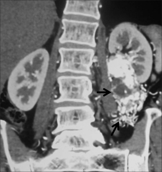
With the ever increasing trend of using cross-section imaging in today's era, incidental detection of small solid renal masses has dramatically multiplied. Coincidentally, the number of asymptomatic benign lesions being detected has also increased. The role of radiologists is not only to identify these lesions, but also go a one step further and accurately characterize various renal masses. Earlier detection of small renal cell carcinomas means identifying at the initial stage which has an impact on prognosis, patient management and healthcare costs. In this review article we share our experience with the typical and atypical solid renal masses encountered in adults in routine daily practice.

Keywords: Adults; RCC; renal masses; solid.


Conflict of interest statement

There are no conflicts of interest.

Figures

Figure 1
Figure 1
Line diagram showing ball and bean-type renal mass
Figure 2 (A and B)
Figure 2 (A and B)
(A and B) Clear cell RCC: Contrast-enhanced CT showing typical features of clear cell RCC –ball type lesion causing smooth contour bulge, showing heterogeneous enhancement with areas of necrosis within the tumor
Figure 3 (A and B)
Figure 3 (A and B)
Necrotic RCC: (A) Contrast-enhanced CT image showing a predominantly necrotic mass in right kidney (B) Axial T2-weighted MR images showing T2 hyperintense necrotic centre with solid component within the tumor
Figure 4 (A and B)
Figure 4 (A and B)
(A, B) Clear cell RCC with calcification: Axial contrast-enhanced CT images showing heterogeneously enhancing ball type lesion in right kidney with specks of calcification within the tumor
Figure 5
Figure 5
Clear cell RCC with paraspinal invasion: Axial contrast-enhanced CT image showing right RCC with extension and invasion into the right paraspinal muscle (dashed arrow)
Figure 6 (A and B)
Figure 6 (A and B)
(A, B) Clear cell RCC in horseshoe kidney: Axial contrast-enhancedCT image showing horseshoe kidneys with a heterogeneously enhancing clear cell RCC in right moiety
Figure 7 (A and B)
Figure 7 (A and B)
(A, B) Multifocal RCC: Coronal CECT images showing two RCCs – one in interpolar region (arrow) and another in lower pole (dashed arrows) in right kidney
Figure 8
Figure 8
Bilateral RCC: Axial contrast-enhancedCT image showing heterogeneously enhancing renal masses in both the kidneys proven to be bilateral RCCs
Figure 9
Figure 9
Papillary RCC: Axial contrast-enhancedCT images showing hypovascular bean-type left renal mass proven to be papillary RCC
Figure 10
Figure 10
Chromophobe RCC: Axial contrast-enhancedCT image showing ball-type left renal mass with spoke-wheel enhancement histopathologically confirmed chromophobe RCC
Figure 11 (A-C)
Figure 11 (A-C)
Medullary RCC:(A, B) Axial contrast-enhancedCT abdomen images showing bean-type right renal mass centrally located (arrow) with right renal vein and IVC tumoral thrombus (dashed arrow) with liver metastases (arrowhead) (C) CT lung section showing pulmonary metastases
Figure 12
Figure 12
Multicystic RCC: Coronal contrast-enhancedCT image showing multicystic upper pole right renal mass with thick enhancing septae and irregular walls suggestive of Bosniak IV cyst, histopathologically proved to be multicystic RCC
Figure 13 (A and B)
Figure 13 (A and B)
Cystic RCC with calcification (A, B): Axial contrast-enhancedCT image showing predominantly cystic lesion with enhancing solid component (arrow) and calcification (arrowhead)
Figure 14
Figure 14
Oncocytoma with central scar: Axial contrast-enhancedCT image showing large left renal mass with central scar (arrowhead) histopathologically proven giant oncocytoma
Figure 15 (A and B)
Figure 15 (A and B)
(A, B) Oncocytoma with segmental enhancement inversion: Coronal contrast- enhanced MR images (A) small ball-type renal mass in lower pole of right kidney showing peripheral enhancement in corticomedullary phase and (B) central enhancement in excretory phase suggestive of oncocytoma
Figure 16
Figure 16
Infiltrative lymphoma: Axial contrast-enhancedCT image showing bilateral enlarged hypodense kidneys (arrow) giving the appearance of bean-type renal mass with enlarged retroperitoneal lymph nodes
Figure 17
Figure 17
Lymphoma –Direct extension from retroperitoneum: Axial contrast-enhancedCT image showing retroperitoneal lymphadenopathy (arrow) infiltrating the left kidney (dashed arrow)
Figure 18
Figure 18
Perinephric lymphoma: Axial contrast-enhancedCT image showing hypodense perinephric soft tissue surrounding the left kidney in a case of perinephric lymphoma
Figure 19
Figure 19
Solitary mass forming lymphoma: Axial contrast-enhancedCT image showing solitary hypoenhancing lesion in left kidney histopathologically confirmed lymphoma
Figure 20
Figure 20
Triphasic angiomyolipoma: Axial contrast-enhancedCT images showing ball-type left renal mass with fat, blood vessel, and soft tissue component
Figure 21 (A and B)
Figure 21 (A and B)
AML in Tuberous sclerosis: (A) CT head image showing calcified subependymal nodules (B) contrast-enhancedCT abdomen image showing multiple fat-containing lesions in bilateral kidneys suggestive of AMLs
Figure 22
Figure 22
Renal metastases: Axial contrast-enhancedCT image showing hypodense multiple metastases in liver, kidneys, and spleen in a known case of carcinoma rectum
Figure 23
Figure 23
Renal metastases: Axial contrast-enhancedCT images showing retroperitoneal lymphadenopathy infiltrating into left kidney in a known case of metastases from testicular malignancy
Figure 24
Figure 24
Infiltrative RCC: Axial contrast-enhancedCT image showing heterogeneously enhancing infiltrative RCC (arrow) in left kidney giving a bean-type appearance with enhancing tumoral thrombus in left renal vein (dashed arrow), and IVC (arrowhead)
Figure 25 (A and B)
Figure 25 (A and B)
(A, B) Infiltrative RCC: Axial CECT images showing expansive bean-type renal mass involving the left kidney (arrow) with retroperitoneal lymphadenopathy
Figure 26
Figure 26
(A, B) Infiltrative TCC: Axial contrast-enhancedCT images showing mildly enhancing bean-type renal mass involving the collecting system extending into the renal pelvis of left kidney (arrows)
Figure 27
Figure 27
Mesenchymal tumor: Coronal contrast-enhancedCT image showing ball-type renal mass (arrow) in left kidney with calcification histopathologically proven to be mesenchymal renal mass
Figure 28 (A and B)
Figure 28 (A and B)
(A, B): Extraosseous Ewing's sarcoma: (A) Unenhanced CT image showing expansive left renal mass with hyperdense contents suggestive of hemorrhage (B) contrast-enhancedCT image showing mild heterogeneous enhancement of the lesion with necrosis histopathologically proven to be extraosseous Ewing's sarcoma of kidney in a 36-year-old-male
Figure 29
Figure 29
Renal lymphangiectasia: Axial contrast-enhancedCT image showing cystic attenuation lesion in bilateral perinephric space suggestive of lymphangiectasia
Figure 30
Figure 30
Renal hemangioma: Axial contrast-enhancedCT image showing intensely enhancing mass (arrow) in left kidney confirmed to be a case of renal hemangioma
Figure 31 (A and B)
Figure 31 (A and B)
(A, B): Cystic nephroma: Coronal T1 and T2-weighted MR images showing multicystic lesion in left kidney with thin septations proven to be cystic nephroma
Figure 32
Figure 32
Renal artery aneurysm: Axial contrast-enhancedCT image showing intensely enhancing left renal artery aneurysm (arrow)
Figure 33
Figure 33
Arteriovenous malformation: Coronal contrast-enhancedCT image showing arteriovenous malformation (arrow) in the left perinephric and pararenal space

Comment in

  • Author reply.
    Indiran V. Indiran V. Indian J Radiol Imaging. 2017 Apr-Jun;27(2):259-260. doi: 10.4103/ijri.IJRI_506_16. Indian J Radiol Imaging. 2017. PMID: 28744091 Free PMC article. No abstract available.

References

    1. Jemal A, Siegel R, Xu J, Ward E. Cancer statistics, 2010. CA Cancer J. 2010;60:277–300. - PubMed
    1. Agnihotri S, Kumar J, Jain M, Kapoor R, Mandhani A. Renal cell carcinoma in India demonstrates early age of onset and a late stage of presentation. Indian J Med Res. 2014;140:624–9. - PMC - PubMed
    1. Kovacs G, Akhtar M, Beckwith BJ, Bugert P, Cooper CS, Delahunt B, et al. The Heidelberg classification of renal cell tumours. J Pathol. 1997;183:131–3. - PubMed
    1. Pahernik S, Ziegler S, Roos F, Melchior SW, Thüroff JW. Small renal tumors: Correlation of clinical and pathological features with tumor size. J Urol. 2007;178:414–7. - PubMed
    1. Gerst S, Hann LE, Li D, Gonen M, Tickoo S, Sohn MJ, et al. Evaluation of renal masses with contrast-enhanced ultrasound: Initial experience. AJR Am J Roentgenol. 2011;197:897–906. - PMC - PubMed