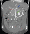
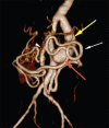
Dacron graft aneurysm with dissection
- PMID: 28104941
- PMCID: PMC5201077
- DOI: 10.4103/0971-3026.195783
Dacron graft aneurysm with dissection
Abstract
Dacron grafts have been used as a conduit for large caliber arteries for many years successfully. However, these grafts can undergo complications such as aneurysm formation, rupture, and failure. Evaluation of these complications are of paramount importance because of its tendency to rupture and cause death. Imaging plays an important role in identifying and monitoring of these complications, and also provides a road map to the vascular surgeons for early intervention and revascularization.
Keywords: Aneurysm; aorta; dacron graft; dissection.
Conflict of interest statement
There are no conflicts of interest.
Figures


References
-
- Noorani A, Ng C, Gopalan D, Dunning J. Haemoptysis from a Dacron graft aneurysm 21 years post repair of coarctation of the aorta. Interact Cardiovasc Thorac Surg. 2011;13:91–3. - PubMed
-
- Nagano N, Cartier R, Zigras T, Mongrain R, Leask RL. Mechanical properties and microscopic findings of a Dacron graft explanted 27 years after coarctation repair. J Thorac Cardiovasc Surg. 2007;134:1577–8. - PubMed
-
- Etz CD, Homann T, Silovitz D, Bodian CA, Luehr M, Di Luozzo G, et al. Vascular graft replacement of the ascending and descending aorta: Do Dacron grafts grow? Ann Thorac Surg. 2007;84:1206–13. - PubMed
Publication types
LinkOut - more resources
Full Text Sources
Other Literature Sources

