

Spontaneous rupture of hepatic epithelioid hemangioendothelioma: A case report
- PMID: 28104995
- PMCID: PMC5221283
- DOI: 10.3748/wjg.v23.i1.185
Spontaneous rupture of hepatic epithelioid hemangioendothelioma: A case report
Abstract
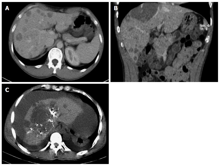
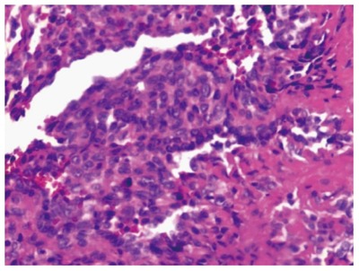
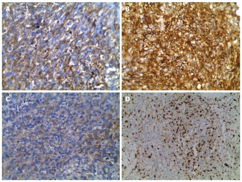
Hepatic epithelioid hemangioendothelioma (HEH) is a rare tumor of vascular endothelial origin. Spontaneous rupture of HEH is a life-threatening complication and is extremely rare. HEH has variable malignant potential, and the clinical diagnosis remains challenging. Here we report a case of HEH with spontaneous rupture. A 44-year-old man presented with constant cutting pains over the right upper abdomen after eating. He had hemoptysis 11 d previously. Diagnostic abdominal puncture demonstrated active bleeding. Chest and abdominal computer tomography scan showed multiple ground-glass nodules over the lungs, multiple low-density intrahepatic nodules and massive hemorrhage. Transcatheter arterial embolization and exploratory laparotomy were performed and subsequent immunohistochemical examination confirmed a diagnosis of HEH.
Keywords: Hepatic epithelioid hemangioendothelioma; Spontaneous rupture.
Figures
Similar articles
-
[A case of primary hepatic epithelioid hemangioendothelioma with spontaneous rupture].Korean J Hepatol. 2009 Dec;15(4):510-6. doi: 10.3350/kjhep.2009.15.4.510. Korean J Hepatol. 2009. PMID: 20037270 Korean.
-
Malignant hepatic epithelioid hemangioendothelioma with abdominal pain due to rapid progression.J Nippon Med Sch. 2011;78(4):246-51. doi: 10.1272/jnms.78.246. J Nippon Med Sch. 2011. PMID: 21869559
-
[A case of hepatic epithelioid hemangioendothelioma that caused extrahepatic metastases without intrahepatic recurrence after hepatic resection].Korean J Hepatol. 2008 Dec;14(4):525-31. doi: 10.3350/kjhep.2008.14.4.525. Korean J Hepatol. 2008. PMID: 19119248 Korean.
-
Hepatic epithelioid hemangioendothelioma: Pitfalls in the diagnosis on fine needle cytology and "small biopsy" and review of the literature.Pathol Res Pract. 2015 Sep;211(9):702-5. doi: 10.1016/j.prp.2015.06.009. Epub 2015 Jul 2. Pathol Res Pract. 2015. PMID: 26187370 Review.
-
Primary malignant hepatic epithelioid hemangioendothelioma: a comprehensive review of the literature with emphasis on the surgical therapy.Cancer. 2006 Nov 1;107(9):2108-21. doi: 10.1002/cncr.22225. Cancer. 2006. PMID: 17019735 Review.
Cited by
-
Hepatic epithelioid hemangioendothelioma: Update on diagnosis and therapy.World J Clin Cases. 2020 Sep 26;8(18):3978-3987. doi: 10.12998/wjcc.v8.i18.3978. World J Clin Cases. 2020. PMID: 33024754 Free PMC article. Review.
-
Three cases of hepatic epithelioid hemangioendothelioma evaluated using conventional and contrast-enhanced ultrasound: Case reports.J Clin Ultrasound. 2022 Jul;50(6):826-831. doi: 10.1002/jcu.23141. Epub 2022 Jan 12. J Clin Ultrasound. 2022. PMID: 35018654 Free PMC article.
-
Spontaneous hepatic haemangioma rupture and hemoperitoneum: a double problem with a single stage interventional radiology solution.Clin Exp Emerg Med. 2019 Jun;6(2):169-172. doi: 10.15441/ceem.18.008. Epub 2019 May 7. Clin Exp Emerg Med. 2019. PMID: 31036786 Free PMC article.
-
Laparoscopic resection of hepatic epithelioid hemangioendothelioma: report of eleven rare cases and literature review.World J Surg Oncol. 2020 Oct 29;18(1):282. doi: 10.1186/s12957-020-02034-z. World J Surg Oncol. 2020. PMID: 33121478 Free PMC article. Review.
-
Vascular tumors in infants and adolescents.Insights Imaging. 2019 Mar 13;10(1):30. doi: 10.1186/s13244-019-0718-6. Insights Imaging. 2019. PMID: 30868300 Free PMC article. Review.
References
-
- Mehrabi A, Kashfi A, Fonouni H, Schemmer P, Schmied BM, Hallscheidt P, Schirmacher P, Weitz J, Friess H, Buchler MW, et al. Primary malignant hepatic epithelioid hemangioendothelioma: a comprehensive review of the literature with emphasis on the surgical therapy. Cancer. 2006;107:2108–2121. - PubMed
-
- Ishak KG, Sesterhenn IA, Goodman ZD, Rabin L, Stromeyer FW. Epithelioid hemangioendothelioma of the liver: a clinicopathologic and follow-up study of 32 cases. Hum Pathol. 1984;15:839–852. - PubMed
-
- Makhlouf HR, Ishak KG, Goodman ZD. Epithelioid hemangioendothelioma of the liver. Cancer. 1999;85:562–582. - PubMed
-
- Earnest F, Johnson CD. Case 96: Hepatic epithelioid hemangioendothelioma. Radiology. 2006;240:295–298. - PubMed
-
- Azzam RI, Alshak NS, Pham HP. AIRP best cases in radiologic-pathologic correlation: Hepatic epithelioid hemangioendothelioma. Radiographics. 2012;32:789–794. - PubMed
Publication types
MeSH terms
Substances
LinkOut - more resources
Full Text Sources
Other Literature Sources
Medical
