Overexpression of Thy1 and ITGA6 is associated with invasion, metastasis and poor prognosis in human gallbladder carcinoma
- PMID: 28105220
- PMCID: PMC5228576
- DOI: 10.3892/ol.2016.5341
Overexpression of Thy1 and ITGA6 is associated with invasion, metastasis and poor prognosis in human gallbladder carcinoma
Abstract
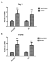
Gallbladder cancer (GBC) is a rare but highly aggressive cancer for which no well-accepted prognostic biomarkers have been identified. Thymus cell antigen 1 (Thy1), also known as cluster of differentiation (CD)90, and integrin α6 (ITGA6), also known as CD49f, are important molecules in cancer and putative markers of various stem cell types. However, their role in GBC remains to be elucidated. In the present study, Thy1 and ITGA6 expression status in clinical GBC samples, which comprised squamous cell/adenosquamous carcinoma (SC/ASC) and adenocarcinoma (AC) subtypes, was investigated. The associations between Thy1 and ITGA6 expression and clinical parameters and survival rate were analyzed separately. The THY1 and ITGA6 messenger RNA levels were significantly higher in both SC/ASC and AC tissues than in adjacent non-tumor tissues (all P<0.001). These results were subsequently confirmed by immunohistochemical analyses. Overexpression of Thy1 and ITGA6 was correlated with poor differentiation, large tumor size, lymph node metastasis and great invasiveness in SC/ASC (Thy1, P=0.045, P=0.005, P=0.003 and P=0.009, respectively, and ITGA6, P=0.029, P=0.011, P=0.009 and P=0.004, respectively) and AC (Thy1, P=0.027, P<0.001, P=0.003 and P 0.004, respectively, and ITGA6, P=0.002, P=0.003, P=0.006 and P=0.006, respectively). Both Thy1 and ITGA6 were expressed at higher levels in AC with advanced tumor-node-metastasis stage (TNM) than in AC with low TNM stage (P=0.001 and P=0.018, respectively). In addition, patients with elevated Thy1 or ITGA6 expression had shorter overall survival than those with negative Thy1 or ITGA6 expression. Multivariate Cox regression analysis demonstrated that Thy1 (SC/ASC, P=0.001 and AC, P=0.005) and ITGA6 (both P=0.003) were independent predictors of poor prognosis in both SC/ASC and AC patients. In conclusion, Thy1 and ITGA6 could be clinical prognostic markers for GBC.
Keywords: ITGA6; Thy1; gallbladder carcinoma; prognostic markers.
Figures


Similar articles
-
Clinicopathological features and CCT2 and PDIA2 expression in gallbladder squamous/adenosquamous carcinoma and gallbladder adenocarcinoma.World J Surg Oncol. 2013 Jun 19;11:143. doi: 10.1186/1477-7819-11-143. World J Surg Oncol. 2013. PMID: 23782473 Free PMC article.
-
N-cadherin and P-cadherin are biomarkers for invasion, metastasis, and poor prognosis of gallbladder carcinomas.Pathol Res Pract. 2014 Jun;210(6):363-8. doi: 10.1016/j.prp.2014.01.014. Epub 2014 Feb 22. Pathol Res Pract. 2014. PMID: 24636838
-
Comparative study of ROR2 and WNT5a expression in squamous/adenosquamous carcinoma and adenocarcinoma of the gallbladder.World J Gastroenterol. 2017 Apr 14;23(14):2601-2612. doi: 10.3748/wjg.v23.i14.2601. World J Gastroenterol. 2017. PMID: 28465645 Free PMC article.
-
Adenosquamous histology predicts a poor outcome for patients with advanced-stage, but not early-stage, cervical carcinoma.Cancer. 2003 May 1;97(9):2196-202. doi: 10.1002/cncr.11371. Cancer. 2003. PMID: 12712471 Review.
-
Regulation and Functions of α6-Integrin (CD49f) in Cancer Biology.Cancers (Basel). 2023 Jul 2;15(13):3466. doi: 10.3390/cancers15133466. Cancers (Basel). 2023. PMID: 37444576 Free PMC article. Review.
Cited by
-
Integrated analysis of the impact of age on genetic and clinical aspects of hepatocellular carcinoma.Aging (Albany NY). 2018 Aug 20;10(8):2079-2097. doi: 10.18632/aging.101531. Aging (Albany NY). 2018. PMID: 30125264 Free PMC article.
-
A Novel Ferroptosis-Associated Gene Signature to Predict Prognosis in Patients with Uveal Melanoma.Diagnostics (Basel). 2021 Feb 2;11(2):219. doi: 10.3390/diagnostics11020219. Diagnostics (Basel). 2021. PMID: 33540700 Free PMC article.
-
PSMC2/ITGA6 axis plays critical role in the development and progression of hepatocellular carcinoma.Cell Death Discov. 2021 Aug 19;7(1):217. doi: 10.1038/s41420-021-00585-y. Cell Death Discov. 2021. PMID: 34413286 Free PMC article.
-
Prognostic and clinicopathological value of CD90 expression in cancer patients: a systematic review and meta-analysis.Transl Cancer Res. 2021 Jul;10(7):3356-3363. doi: 10.21037/tcr-21-266. Transl Cancer Res. 2021. PMID: 35116641 Free PMC article.
-
Determination of WWOX Function in Modulating Cellular Pathways Activated by AP-2α and AP-2γ Transcription Factors in Bladder Cancer.Cells. 2022 Apr 19;11(9):1382. doi: 10.3390/cells11091382. Cells. 2022. PMID: 35563688 Free PMC article.
References
LinkOut - more resources
Full Text Sources
Other Literature Sources
Miscellaneous
